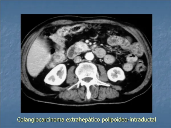
Colangiocarcinoma extrahepático polipoideo-intraductal
140 likes | 1k Vues
Colangiocarcinoma extrahepático polipoideo-intraductal. Colangiocarcinoma intrahepático polipoideo-intraductal. Colangiocarcinoma extrahepático polipoideo-intraductal: plano radial SSFSE con TR 2528 y TE 1477.1, de grosor 50.0.

Colangiocarcinoma extrahepático polipoideo-intraductal
E N D
Presentation Transcript
Colangiocarcinoma extrahepático polipoideo-intraductal: plano radial SSFSE con TR 2528 y TE 1477.1, de grosor 50.0
Colangiocarcinoma extrahepático polipoideo-intraductal: plano coronal SSFSE con TR 1910 y TE 119.0, de grosor 7.0
Colangiocarcinoma extrahepático polipoideo-intraductal: plano axial SSFSE con TR 1910 y TE 120.6, de grosor 3.0
Colangiocarcinoma extrahepático polipoideo-intraductal: plano axial SSFSE con TR 1910 y TE 120.6, de grosor 3.0
Colangiocarcinoma extrahepático polipoideo-intraductal: plano radial SSFSE con TR 2574 y TE 1431.0, de grosor 50.0
CONCLUSIONES • Distribución ligeramente mayor en varones. • Localización hiliar la más frecuente. • TC > 90%, RM casi 50%. • AP adenocarcinoma en 96%. • Técnicas de elección para estudio: TC y RM para tumores intrahepáticos , ERCP y colangioRM para extrahepáticos.
AGRADECIMIENTOS Al servicio de Anatomía Patológica del H. U. Miguel Servet de Zaragoza, en particular a la Dra. Ana Fuertes, por su colaboración con las imágenes de las piezas quirúrgicas
BIBLIOGRAFÍA • 1. Jae Hoon Lim. Cholangiocarcinoma: Morphologic classification according to growth pattern and imaging findings. AJR 2003; 181:819-27. • 2. Slattery JM, Sahani DV. What is the current state of the art imaging for detection and staging of cholangiocarcinoma? Oncologist 2006; 11: 913-22. • 3. Bismuth H, Majno PE. Biliary strictures: classification based on the principles of surgical treatment. World J Surg 2001; 25: 1241- 44. • 4. Lee HY, Kim SH, Lee JM, Kim SW, Jang JY, Han JK, Choi BI. Preoperative assessment of resectability of hepatic hilar cholangiocarcinoma: combined CT and cholangiography with revised criteria. Radiology. 2006 Apr;239(1):113-21. • 5. Asayama Y, Yoshimitsu K, Irie H, Tajima T, Nishie A, Hirakawa M, Nakayama T, Kakihara D, Taketomi A, Aishima S, Honda H.Delayed-phase dynamic CT enhancement as a prognostic factor for mass-forming intrahepatic cholangiocarcinoma. Radiology. 2006 Jan;238(1):150-5.
6. Joon Koo Han, Byung Ihn Choi, Ah Young Kim, Su Kyung An, Joon Woo Lee, Tae Kyung Kim, and Sun-Whe Kim. Cholangiocarcinoma: Pictorial Essay of CT and Cholangiographic Findings. RadioGraphics 2002; 22: 173-187. • 7. Yuji Watanabe, Masako Nagayama, Akira Okumura, Yoshiki Amoh, Takashi Katsube, Tsuyoshi Suga, Shingo Koyama, Kohya Nakatani, and Yoshihiro Dodo. MR Imaging of Acute Biliary Disorders. RadioGraphics 2007; 27: 477-495. • 8. Jae Hoon Lim, Kee-Taek Jang, Dongil Choi, Won Jae Lee, and Hyo Keun Lim. Early Bile Duct Carcinoma: Comparison of Imaging Features with Pathologic Findings. Radiology 2006 238: 542-548. • 9. Mi-Suk Park, Tae Kyoung Kim, Kyyoung Won Kim, Sung Won Park, Jeong Kyung Lee, Jung-Sun Kim et al. Differentiation of extrahepatic bile duct cholangiocarcinoma from benign stricture: findings at MRCP versus ERCP. Radiology 2004; 233: 234-240.