Download

1 / 20
200 likes | 546 Vues
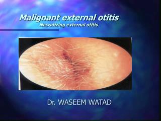
Malignant external otitis Necrotizing external otitis. Dr. WASEEM WATAD. Case 1. ( SH. Y ). 80 years old 3VD , PTCA , DM-type2 , HTN , BPH Ext. otitis with PO ABX and ear drops with improvement several months before admission

E N D
Malignant external otitis Necrotizing external otitis Dr. WASEEM WATAD
Case 1. ( SH. Y ) • 80 years old • 3VD , PTCA , DM-type2 , HTN , BPH • Ext. otitis with PO ABX and ear drops with improvement several months before admission • severe Rt. otalgia , facial pain Rt. , and Rt. parotid mass at admission 19/09/04 • Rt ear discharge • Weight loss
Case 1. • CT scan (20/09/04): Rt parotid mass , infiltration of parapharyngeal fat , EAC , infratemporal fossa , Rt. lat. pterygoid and masseter .no bony erosion and no lymphadenopathy • MRI (19/10/04) :process infiltrating the Rt. ear,temporal bone , TMJ, sphenoid sinus , infratemporal fossa and skull base • Biopsy of EAC polyp, parotid FNA (28/10/04) – mixed inflammation • Positive culture for p. aeruginosa
Case 1. • IV ABX treatment ( cephalosporine and quinolones ) with ear drops and toilette • Improvement in pain , ear discharge • There was no CN involvement
Case 2. ( Va. D ) • 68 years old • DM-type 2 , HTN • Hyperlipidemia , s/p CVA • Rt. Nasopharyngeal mass – biopsy no malignancy (11/04) • Bil. Ext. otitis 09/04 ( several weeks before admittion ) prolong ABX treatment ( semi-synthetic penicillin , quinolone) and ear drops
Case 2. • No improvement • Rt. Severe otalgia , ear discharge , persistent rt. ext. otitis , with granulation tissue • Elevated ESR , negative culture for p. aeruginosa • Start IV ceftazidime ( 5 weeks ) • Progression findings in serial CT/MRI
Case 2. • CT scan ( 14/11/04 ) - infiltration of the rt. parapharyngeal space , rt. Mastoid and middle ear, infiltrating of infratemporal fossa • MRI ( 24/21/04 ) – large mass in rt. parapharyngeal space with involvement of rt. TMJ and deep lobe of rt. Parotis • CT (01/05) infiltrating in rt. TMJ
Case 2. • De’bridment - (10/01/05) ,. (24/01/05), • Hx – inflammatory tissue • 2 weeks of AMIKACIN + MEROPENEM • Exacerbation of Rt. Otalgia , ear discharge and relapse of granulation tissue of EAC • Treatment failure ?? • Further therapy : • Broad spectrum of ABX – combination of cephalosporines and quinolone • Surgical treatment – mastoidectomy +/- tympanoplasty , ablation of granulating and necrotizing tissue, bone and cartilage sequestrations • HBO
Parietal Frontal Temporal Sphenoid Z Maxilla Lat. Pterygoid Plate Pterygomaxillary Fissure Infratemporal Fossa
MEO - criteria • Sade’ (1989) : • Severe EXT. otitis unresponsive to at least 10 days of conservative treatment • Increasing agonizing pain exacerbated at night • Granulation tissue in the base of EAC • Repeated isolation of pseudomonas • Levenson (1991) : • Refractory otitis ext. • Severe otalgia , worse at night • Purulent exudate , granulation tissue • Recovery of P. aeruginosa • DM , immune state compromise • Positive Tc-99 bone scan of temporal bone
MEO - staging • Corey (1985) : • I - Infection of bone and soft tissue without cranial nerves lesions or intracranial lesions • II- cranial nerve paralysis • a- VII paralysis only • b- Multiple cranial nerves paralysis • III – meningitis , epidural empyema , subdural empyema or brain abscess
NEO - diagnosis • Clinical findings • Laboratory tests • Culture • Ga-67, Tc-99 scans • HR-CT with contrast • Biopsy of granulation tissue
mortality • 46% (1968) • 10% recent articles • High mortality in facial n. paralysis
Management – cont. • HR-CT contrast evaluation • Ga-67 (every 4 weeks) follow up with treatment • Management underlying process ( DM / immunosuppressive) • Surgical de’bridment ,drinage – intracranial ext. , brain abscess • 6 weeks of ABX , repeat cultures , oral ABX after 2 weeks of cessation of symptoms
Management – cont. • Deep biopsy of granulation tissue – underlying carcinoma
Therapeutic problems • Main problem is : • Choice of the ABX • Duration of treatment
Therapeutic problems • Duration of treatment • Standard indication ( 6-8 weeks ) • Identifying objective parameter of definitive recovery • Healing of skin EAC • ESR • Ga-67
Therapeutic problems • Surgical treatment : • Complementary role • Mastoidectomy +/- tympanoplasty • Recommendation – biopsy , cleansing , ablation of necrotizing and granulation tissue and the bone , cartilage sequestrations
Therapeutic problems • Hyperbaric oxygen therapy • Daily , 2.4-3 atm, 90 minutea , 30 courses • Indications : advanced stages , recurrent cases, refractory to ABX • Hypoxia impaired oxygen dependent bacterial killing by phagocytosis