Download
1 / 11
110 likes | 271 Vues
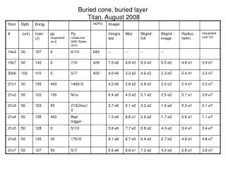
Buried cone, buried layer Titan, August 2008. Buried cone buried layer comments. Is it possible laser didn’t hit bottom every time? Intrinsic prepulse -- 19s7, 21s5 - the latter has very well defined peak. Former much broader.

E N D
Buried cone buried layer comments • Is it possible laser didn’t hit bottom every time? • Intrinsic prepulse -- 19s7, 21s5 - the latter has very well defined peak. Former much broader. • 50 mJ prepulse -- 21s3, 21s6, 21s7 have -- peak definition improves with later shots. • Some of the well-defined spots weren‘t in the center of the foil. The foils don’t seem to be offset that much - see 21s5,7. Need to work out relationship of x-radiographs to target images to check. • Foil is not much larger than peak - but checked measuring routine against lineouts, and find results OK.
19s7 Bur 50 m #1 • Target #1 • Image blurred by 30 pixels for peak measurement. • Foil 350 m across => 1 pixel = 3 m • Images 460 pixels across • Lineout suggests bkgnd 8 e2 FWHM X-radiograph 5 pixel blur 30 pixel blur Profile
20s6 Bur 100 m #1 • Image blurred by 30 pixels for peak measurement. • Foil 350 m across => 1 pixel = 3 m • Images 460 pixels across • Edge is noticeably lit up, but doesn’t have a significant number of counts • Linout suggests bkgnd 5.5 e2 FWHM X-radiograph 5 pixel blur 30 pixel blur Profile
21s1 Bur 50 m #2 • Image blurred by 30 pixels for peak measurement. • Foil 350 m across => 1 pixel = 3 m • Images 460 pixels across • Lineout suggests 5 e2 bkgnd FWHM X-radiograph 5 pixel blur 30 pixel blur Profile
21s2 Bur 50 m #3 • Image blurred by 30 pixels for peak measurement. • Foil 350 m across => 1 pixel = 3 m • Images 460 pixels across • Lineout suggests bkgnd 5 e2 FWHM X-radiograph 5 pixel blur 30 pixel blur Profile
21s3 Bur 50 m #4 • Image blurred by 30 pixels for peak measurement. • Foil 350 m across => 1 pixel = 3 m • Images 460 pixels across • Lineout suggests bkgnd 3 e2 FWHM X-radiograph 5 pixel blur 30 pixel blur Profile
21s4 Bur 50 m #5 • Image blurred by 30 pixels for peak measurement. • Foil 350 m across => 1 pixel = 3 m • Images 460 pixels across • Lineout shows 3 e2 foil intensity FWHM X-radiograph 5 pixel blur 30 pixel blur Profile
21s5 Bur 50 m #6 • Image blurred by 30 pixels for peak measurement. • Foil 350 m across => 1 pixel = 3 m • Images 460 pixels across • Lineout suggets bkgnd 7 e2 FWHM X-radiograph 5 pixel blur 30 pixel blur Profile
21s6 Bur 50 m #7 • Image blurred by 30 pixels for peak measurement. • Foil 350 m across => 1 pixel = 3 m • Images 460 pixels across • Lineout suggests bkgnd 6.5 e2 FWHM X-radiograph 5 pixel blur 30 pixel blur Profile
21s7 Bur 50 m #8 • Image blurred by 30 pixels for peak measurement. • Foil 350 m across => 1 pixel = 3 m • Images 460 pixels across • Lineout suggests bkgnd 7 e2 FWHM X-radiograph 5 pixel blur 30 pixel blur Profile