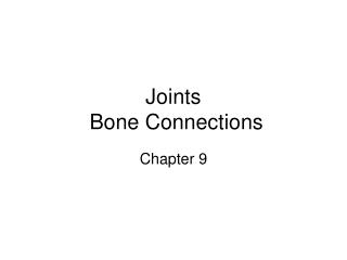
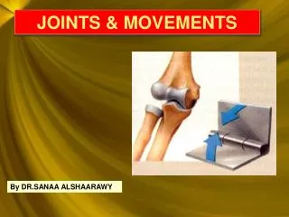
JOINTS & MOVEMENTS
JOINTS & MOVEMENTS. By DR.SANAA ALSHAARAWY. Objectives. By the end of the lecture, the student should be able to : List the functional & structural classification of the joints. Name the 3 different types of joints. Describe the structure of each type. Name an example of each type.

JOINTS & MOVEMENTS
E N D
Presentation Transcript
JOINTS & MOVEMENTS By DR.SANAA ALSHAARAWY
Objectives • By the end of the lecture, the student should be able to: • List the functional & structural classification of the joints. • Name the 3 different types of joints. • Describe the structure of each type. • Name an example of each type. • Describe the movements which occur in each type.
WHAT IS A JOINT ? INTRODUCTION It is a meeting of two or more bones. • Every bone in the body forms ajointwith at least one or more bone, (except the hyoid bone in the neck). • Joints have two functions: • They hold the bones together securely. • Also it give the rigid skeleton mobility. Femur Patella Tibia Fibula X-ray of the knee joint
CLASSIFICATION Joints could be classified by two ways: • According to function Or • According to structure.
I- FUNCTIONAL CLASSIFICATION The functional classification depends on the amount of movement allowed by the joint. On this basis, there are: • Synarthroses or immovable joints: Skull sutures. 1- Synarthroses or immovable joints, 2- Amphiarthroses or slightly movable joints. 3- Diarthrosesor freely movable joints.
Amphiarthroses or slightly movable joints, e.g. Symphysis pubis.
Immovable & slightly movable jointsare restricted mainly to the axial skeleton,where firm attachments and protection of internal organs are priorities. • Freely movable joints predominate in the limbs, where mobility is important.
II- STRUCTURAL CLASSIFICATION • This classification depends upon the tissue which connects the bones. • There are three types of joints: • A. Fibrous, • B. Cartilage, or, • C. Synovial joint, where a joint cavity separates the bones. • They are called: • Fibrous joints: Skull sutures Prof. Saeed Makarem
AS A GENERAL RULE: fibrous jointsareimmovable synovial jointsare freely movable most cartilaginous joints are slightly movable(amphiarthroses)
FIBROUS JOINTS • Infibrous joints,the bones are united by fibrous tissue. • Examples: • A- Skull sutures • In this joint, the irregular edges of bones interlock and are bound together tightly by fibers tissue, where no movement are allowed.
FIBROUS JOINTS • B- Inferior tibiofibular joint, no or very minimal movement is allowed. • It is called syndesmoses.
CARTILAGINOUS JOINTS Incartilaginous joints,the 2 bone ends are connected by cartilage. Examples: • Pubic symphysisof the pelvis (slightly movable(amphiarthroses)
CARTILAGINOUS JOINTS • The intervertebral discs of the vertebral column, where the articulating bone surfaces are connected by pads (discs) of fibrocartilage, are alsoslightly movable (amphiarthroses) .
Thehyaline-cartilageEpiphysial plates of growing long bones are immovable (synarthroses) cartilaginous joints. REMEBER !
The cartilaginous jointsbetween the first ribs and the sternumare also immovable (synarthroses)cartilaginous joints. REMEBER !
SYNOVIAL JOINTS • Synovial jointsare those in which the articulating bone ends are separated by a joint cavity which contains a synovial fluid. • They account for all joints of the limbs.
FEATURES OF SYNOVIAL JOINTS • Articular cartilage. The 2 ends of the bones are covered by articular (hyaline) cartilage. 1- Articular cartilage 2- Fibrous capsule 3- Synovial membrane 4- Joint cavity 5- Extracapsular ligaments
Fibrous capsule. The joint surfaces are enclosed by a capsule of fibrous tissue & 3. The capsule is lined with a synovial membrane
4. Joint cavity • Inside the synovial membrane there is a lubricating (synovial fluid).
5. Reinforcing ligaments. The fibrous capsule is usually reinforced with ligaments. (extracapsular ligaments).
TYPES OF SYNOVIAL JOINTS BASED ON SHAPE • Based on the shape, the synovial joints can be classified as: • Plane, • Hinge, • Pivot, • Condyloid, • Saddle, • Ball and socket joint.
PLANE JOINTS • In aplane joint, the articular surfaces are flat, and only short slipping or gliding movements are allowed. • Example: The intercarpal joints of the wrist.
HINGE JOINTS • In ahinge joint, the cylindrical end of one bone fits into a trough-shaped surface on another bone. • Movement is allowed in just one plane, like a hinge. • Hinge joints are Uniaxial ; they allow movement around one axis only. • Examples: elbow & ankle joints, the interphalangeal joints of the fingers. Axis of movement
PIVOT JOINT • In a pivot joint, the rounded end of one bone fits into a ring of bone (or ligaments). • Because the rotating bone can turn only around its long axis, pivot joints are also Uniaxial joints. • Examples: proximal radioulnar joint Axis of movement
CONDYLOID JOINTS • In a condyloid joint, the egg-shaped articular surface of one bone fits into an oval concavity in another. • Both of these articular surfaces are oval. • Movement occurs around two axes, hence these joints are biaxial, as in knuckle (metacarpophalangeal) joints. • Condyloid joints allow the moving bone to move: • from side to side and • back and forth, • but the bone cannot rotate around its long axis. Axes of movement
SADDLE JOINTS • In saddle joints,each articular surface has both convex and concave areas, like a saddle. • These biaxialjoints allow essentially the same movements as condyloid joints. • Example: carpometacarpal joint of the thumb. Axes of movement
BALL-AND-SOCKET JOINTS • In aball-and-socket joint, the spherical head of one bone fits into a round socket in another. • I t is a multiaxialjoints allow movement in all axes, including rotation, and are the most freely movable joints. • Examples:shoulder and hip.
FLEXION, EXTENSION & HYPEREXTENSION MOVEMENTS More increasing the angle between 2 bones or parts of body. Decreasing angle of joint and brings 2 bones closer together. Increasing angle between 2 bones or parts of the body.
ABDUCTION, ADDUCTION, CIRCUMDUCTION ROTATION A movement of a bone around its longitudinal axis as in ball & socket j.and atlas around dense of axis as in shaking head “No”. Abduction : moving of a limb away from midline,also in fanning of fingers or toes . Adduction : the opposite of abduction, movement of a limb toward body midline.
INVERSION AND EVERSION DORSIFLEXION AND PLANTAR FLEXION Inversion : turning sole of foot medially. Eversion : turning sole of foot laterally. Dorsiflexion : up movement of the foot at ankle j. as standing on heel. Plantar flexion : down movement of foot as in pointing the toes.
SUPINATION AND PRONATION Rotation of forearm laterally so that the palm faces anteriorlly. Rotation of of forearm medially so that the palm faces posteriorly.
OPPOSITION A movement of The thumb to touch the tips of other fingers. It occurs in Saddle joint of 1st Carpo-metacarpal joint of the thumb.