Download
1 / 85
1k likes | 2.27k Vues
The Skeletal System (Bones and Joints). Anatomy & Physiology I Chapter 7. Bone as a Tissue. osteology – the study of bone skeletal system - composed of bones, joints, cartilages, and ligaments form strong flexible framework of the body cartilage – forerunner of most bones

E N D
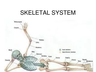
The Skeletal System (Bones and Joints) Anatomy & Physiology I Chapter 7
Bone as a Tissue • osteology – the study of bone • skeletal system - composed of bones, joints, cartilages, and ligaments • form strong flexible framework of the body • cartilage – forerunner of most bones • covers many joint surfaces of mature bone • ligaments – hold bones together at the joints • tendons – attach muscle to bone
Functions of the Skeleton • support – hold the body up, supports muscles, mandible and maxilla support teeth • protection – brain, spinal cord, heart, lungs • movement – limb movements, breathing, action of muscle on bone • electrolyte balance – calcium and phosphate ions • acid-base balance – buffers blood against excessive pH changes • blood formation – red bone marrow is the chief producer of blood cells
Bones and Osseous Tissue • bone (osseous tissue) - connective tissue with the matrix hardened by calcium phosphate and other minerals • mineralization or calcification – the hardening process of bone • individual bones consist of bone tissue, bone marrow, cartilage, adipose tissue, nervous tissue, and fibrous connective tissue • continually remodels itself and interacts physiologically with all of the other organ systems of the body • permeated with nerves and blood vessels, which attests to its sensitivity and metabolic activity
The Matrix of Bone • matrix of osseous tissue is, by dry weight, about one-third organic and two-thirds inorganic matter • organic matter – synthesized by osteoblasts • collagen, carbohydrate – protein complexes, such as proteoglycans and glycoproteins • inorganic matter • 85% calcium phosphate (hydroxyapatite) • 10% calcium carbonate • other minerals (fluoride, sodium, potassium, magnesium) • bone is a composite – combination of two basic structural materials, a ceramic and a polymer • combines optimal mechanical properties of each component • bone combines the polymer, collagen, with the ceramic, hydroxyapatite and other minerals • ceramic portion allows the bone to support the body weight, and protein portion gives bone some degree of flexibility • rickets – soft bones due to deficiency of calcium salts • osteogenesis imperfecta or brittle bone disease – excessively brittle bones due to lack of protein, collagen
Bone Structure Types of osseous (bone) tissue • Compact bone - Dense outer layer • Spongy (cancellous) bone - Honeycomb of trabeculae • Bone marrow • Red marrow • Yellow marrow • Bone membranes • Periosteum • Endosteum
Spongy bone (diploë) Compact bone Trabeculae
Structure of a Long Bone • Diaphysis (shaft) • Compact bone collar surrounds medullary (marrow) cavity • Medullary cavity in adults contains fat (yellow marrow) • Epiphyses • Expanded ends • Spongy bone interior • Epiphyseal line (remnant of growth plate) • Articular (hyaline) cartilage on joint surfaces Articular cartilage Epiphysis Red bone marrow Epiphyseal line Marrow cavity Yellow bone marrow Periosteum Nutrient foramen Diaphysis Compact bone Spongy bone Epiphyseal line Epiphysis Articular cartilage Living Dried
Structure of a Long Bone Articular cartilage Compact bone Proximal epiphysis Spongy bone Epiphyseal line Periosteum Compact bone Medullary cavity (lined by endosteum) Diaphysis Distal epiphysis
Membranes of Bone • Periosteum • Outer fibrous layer • Inner osteogenic layer • Osteoblasts (bone-forming cells) • Osteoclasts (bone-destroying cells) • Osteogenic cells (stem cells) • Nerve fibers, nutrient blood vessels, and lymphatic vessels enter the bone via nutrient foramina • Secured to underlying bone by Sharpey’s fibers
Membranes of Bone • Endosteum • Delicate membrane on internal surfaces of bone • Also contains osteoblasts and osteoclasts
Endosteum Yellow bone marrow Compact bone Periosteum Perforating (Sharpey’s) fibers Nutrient arteries
Structure of Short, Irregular, and Flat Bones • Periosteum-covered compact bone on the outside • Endosteum-covered spongy bone within • Spongy bone called diploë in flat bones • Bone marrow between the trabeculae
Spongy bone (diploë) Compact bone Trabeculae
Bone Marrow • bone marrow – general term for soft tissue that occupies the marrow cavity of a long bone and small spaces amid the trabeculae of spongy bone • red marrow (myeloid tissue) • in nearly every bone in a child • hemopoietic tissue - produces blood cells and is composed of multiple tissues in a delicate, but intricate arrangement that is an organ to itself • in adults, found in skull, vertebrae, ribs, sternum, part of pelvic girdle, and proximal heads of humerus and femur • yellow marrow found in adults • most red marrow turns into fatty yellow marrow • no longer produces blood
Location of Hematopoietic Tissue (Red Marrow) • Red marrow cavities of adults • Trabecular cavities of the heads of the femur and humerus • Trabecular cavities of the diploë of flat bones • Red marrow of newborn infants • Medullary cavities and all spaces in spongy bone
Microscopic Anatomy of Bone • Cells of bones • Osteogenic (osteoprogenitor) cells – give rise to osteoblasts • Stem cells in periosteum, endosteum and central canals • Osteoblasts – manufacture bone matrix • Bone-forming cells • Osteocytes – maintain and repair existing bone matrix • Mature bone cells • Osteoclasts – breakdown (resorb) bone matrix • Cells that break down and release minerals from bone matrix
Microscopic Anatomy of Bone: Compact Bone • Haversian system, or osteon—structural unit • Lamellae • Weight-bearing • Column-like matrix tubes • Central (Haversian) canal • Contains blood vessels and nerves
Microscopic Anatomy of Bone: Compact Bone • Perforating (Volkmann’s) canals • At right angles to the central canal • Connects blood vessels and nerves of the periosteum and central canal • Lacunae—small cavities that contain osteocytes • Canaliculi—hairlike canals that connect lacunae to each other and the central canal
Spongy bone Compact bone Compact Bone Central (Haversian) canal Perforating (Volkmann’s) canal Endosteum lining bony canals and covering trabeculae Osteon (Haversian system) Circumferential lamellae Perforating (Sharpey’s) fibers Periosteal blood vessel Lamellae Periosteum Nerve Vein Lamellae Artery Central canal Lacuna (with osteocyte) Canaliculi Osteocyte in a lacuna Lacunae Interstitial lamellae
Compact Bone Nerve Vein Lamellae Artery Central canal Canaliculus Lacunae Osteocyte in a lacuna
Hormonal Regulation of Bone Growth • Growth hormone stimulates epiphyseal plate activity • Thyroid hormone modulates activity of growth hormone • Testosterone and estrogens (at puberty) • Promote adolescent growth spurts • End growth by inducing epiphyseal plate closure
Bone Deposit • Occurs where bone is injured or added strength is needed • Requires a diet rich in protein; vitamins C, D, and A; calcium; phosphorus; magnesium; and manganese
Bone Resorption • Osteoclasts secrete • Lysosomal enzymes (digest organic matrix) • Acids (convert calcium salts into soluble forms) • Dissolved matrix is transcytosed across osteoclast, enters interstitial fluid and then blood
Control of Remodeling • What controls continual remodeling of bone? • Hormonal mechanisms that maintain calcium homeostasis in the blood • Mechanical and gravitational forces
Hormonal Control of Blood Ca2+ • The level of blood calcium (Ca2+) must be maintained (homeostasis) • Calcium is necessary for • Transmission of nerve impulses • Muscle contraction • Blood coagulation • Secretion by glands and nerve cells • Cell division • Which hormones are responsible for maintaining blood calcium?
Hormonal Control of Blood Ca2+ • Primarily controlled by parathyroid hormone (PTH) Blood Ca2+ levels Parathyroid glands release PTH PTH stimulates osteoclasts to degrade bone matrix and release Ca2+ • Blood Ca2+ levels When blood calcium levels fall, PTH is released causing blood calcium levels to increase.
Hormonal Control of Blood Ca2+ • May be affected to a lesser extent by calcitonin Blood Ca2+ levels Parafollicular cells of thyroid release calcitonin Osteoblasts deposit calcium salts Blood Ca2+ levels When blood calcium levels rise, calcitonin is released causing blood calcium levels to decrease.
Response to Mechanical Stress • Wolff’s law: A bone grows or remodels in response to forces or demands placed upon it • Observations supporting Wolff’s law: • Handedness (right or left handed) results in bone of one upper limb being thicker and stronger • Curved bones are thickest where they are most likely to buckle • Trabeculae form along lines of stress • Large, bony projections occur where heavy, active muscles attach
Bone Markings • Bulges, depressions, and holes serve as • Sites of attachment for muscles, ligaments, and tendons • Joint surfaces • Conduits for blood vessels and nerves
Bone Markings: Projections • Sites of muscle and ligament attachment • Tuberosity—rounded projection • Crest—narrow, prominent ridge • Trochanter—large, blunt, irregular surface • Line—narrow ridge of bone • Tubercle—small rounded projection • Condyle – rounded projection • Epicondyle—raised area above a condyle • Spine—sharp, slender projection • Process—any bony prominence
Bone Markings: Projections • Projections that help to form joints • Head • Bony expansion carried on a narrow neck • Facet • Smooth, nearly flat articular surface • Condyle • Rounded articular projection • Ramus • Armlike bar
Meatus Canal-like passageway Sinus Cavity within a bone Fossa Shallow, basinlike depression Groove Furrow Fissure Narrow, slitlike opening Foramen Round or oval opening through a bone Bone Markings: Depressions and Openings
Bones of the Axial Skeleton Two main groups of bones • Axial skeleton—80 bones of the head and trunk • Appendicular skeleton—126 bones of the extremities
Framework of the Skull • Cranial bones • Facial bones • Infant skull
Framework of the Skull, cont’d • Cranial bones • Frontal • Parietal • Temporal • Ethmoid • Sphenoid • Occipital
Framework of the Skull, cont’d • Facial bones • Mandible • Maxillae • Zygomatic • Nasal • Lacrimal • Vomer • Palatine • Inferior nasal conchae • Ossicle • Hyoid
Framework of the Skull, cont’d • Infant skull • Anterior fontanel
The skull ZOOMING IN • What type of joint is between bones of the skull?
The skull, inferior view. ZOOMING IN • What two bones make up each side of the hard palate?
Floor of cranium, superior view. ZOOMING IN • What is a foramen?
Infant skull, showing fontanels ZOOMING IN • Which is the largest fontanel?
Framework of the Trunk • Vertebral column • Cervical vertebrae • Thoracic vertebrae • Lumbar vertebrae • Sacral vertebrae (sacrum) • Coccygeal vertebrae (coccyx) • Thorax • Sternum • Ribs • True ribs • False ribs • Manubrium • Clavicular notch • Sternal angle • Xiphoid process
The Vertebral Column (Spine) Anterior view Posterior view • five vertebral groups • 7 cervical in the neck • 12 thoracic in the chest • 5 lumbar in lower back • 5 fused sacral at base of spine • 4 fused coccygeal Atlas (C1) Axis (C2) Cervical vertebrae C7 T1 Thoracic vertebrae T12 L1 Lumbar vertebrae L5 S1 Sacrum S5 Coccyx Coccyx