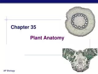
Chapter 35: The Senses
400 likes | 427 Vues
Explore the intricate world of sensory perception and systems, from sensory receptors to somatic sensations, taste, smell, balance, vision, and more. Understand how organisms receive signals, process stimuli, and perceive the external and internal environment.

Chapter 35: The Senses
E N D
Presentation Transcript
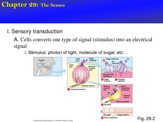
Chapter 35: The Senses Sensory Perception
Sensory Systems • The means by which organisms receive signals from the external world and internal environment • Many animals can sense stimuli that humans cannot
Sensory Receptors Convert the energy of a stimulus into action potentials Mechanoreceptors Thermoreceptors Pain receptors Chemoreceptors Osmoreceptors Photoreceptors
Sensory Pathways d Message travels from stimulated sensory neuron to motor neuron and interneuron in spinal cord c Strectched muscle simulates a stretch receptor (the ending of a sensory neuron) that is adjacent to it. sensory neuron interneuron in spinal cord motor neuron in spinal cord axon endings of motor neuron terminating on the same muscle e Message is sent back to the muscle, also to other interneurons in the brain. muscle spindle Fig. 35-2b, p.601
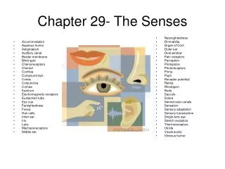
Somatic Sensations • Touch • Pressure • Temperature • Pain • Motion • Position
Somatosensory Cortex Figure 35.4 Page 602
Receptors in Skin • Free nerve ending • Ruffini ending • Pacinian corpuscle • Bulb of Krause • Meissner’s corpuscle Figure 35.5 Page 603
Referred Pain • Sensations of pain from internal organs may be wrongly projected to part of the skin surface • Heart attack can be felt as pain in skin above the heart and along the left shoulder and arm
lungs, diaphragm heart stomach liver, gallbladder pancreas small intestine ovaries colon appendix urinary bladder kidney ureter Fig. 35-6, p.603
Taste • A special sense • Chemoreceptors • Five primary sensations: • sweet, sour, salty, bitter, and umami Figure 35.8 Page 604
Smell • A special sense • Olfactory receptors • Receptor axons lead to olfactory lobe olfactory bulb receptor cell Figure 35.7 Page 604
Balance and Equilibrium • In humans, organs of equilibrium are located in the inner ear • Vestibular apparatus semicircular canals utricle saccule vestibular apparatus Figure 35.9bPage 605
Acceleration-Deceleration • Moving in response to gravity, otoliths bend projections of hair cells and stimulate the endings of sensory neurons otoliths hair cell membrane vestibular nerve
Dynamic Equilibrium • Rotating head movements cause pressure waves that bend a gelatinous cupula and stimulate hair cells inside it cupula
Properties of Sound • Ear detects pressure waves • Amplitude of waves corresponds to perceived loudness • Frequency of waves (number per second) corresponds to perceived pitch
Sound Reception • Sound waves make the eardrum vibrate • Vibrations are transmitted to the bones of the middle ear • The stirrup transmits force to the oval window of the fluid-filled cochlea
scala vestibuli cochlear duct organ of Corti scala tympani sensory neurons (to the auditory nerve) Fig. 35-11d, p.607
Vision • Sensitivity to light does not equal vision • Vision requires two components • Eyes • Capacity for image formation in the brain
Invertebrate Eyes ommatidium Limpet ocellus cuticle epidermis lens Compound eye of a deerfly sensory neuron Figures 35.13 & 35.14Pages 608 & 609 Land snail eye
Invertebrate Eyes Fig. 35-13d, p.608
Invertebrate Eyes Fig. 35-1,e, p.608
vitreous body lens cornea retina optic tract Fig. 35-15, p.609
Human Eye sclera retina choroid iris fovea optic disk lens pupil cornea part of optic nerve aqueous humor ciliary muscle Figure 35.17Page 610 vitreous body
Pattern of Stimulation • Light rays pass through lens and converge on retina at back of eye • The image that forms on the retina is upside down and reversed right to left compared with the stimulus • Brain accounts for this during processing
a Light rays from an object converge on the retina, form an inverted, reversed image. muscle contracted b When a ciliary muscle contracts, the lens bulges, bending the light rays from a close object so that they become focused on the retina. close object slack fibers muscle relaxed c When the muscle relaxes, the lens flattens, focusing light rays from a distant object on the retina. distant object taut fibers Fig. 35-18, p.611
Visual Accommodation • Adjustments of the lens • Ciliary muscle encircles lens • When this muscle relaxes, lens flattens, moves focal point farther back • When it contracts, lens bulges, moves focal point toward front of eye
Organization of Retina • Photoreceptors lie at the back of the retina, in front of a pigmented epithelium • For light to reach the photoreceptors, it must pass layers of neurons involved in visual processing
cone cell stacked, pigmented membrane rod cell Fig. 35-19, p.612
Organization of Retina • Signals from photoreceptors are passed to bipolar sensory neurons, then to ganglion cells Figure 35.20Page 612
The Photoreceptors • Rods • Contain the pigment rhodopsin • Detect very dim light, changes in light intensity • Cones • Three kinds; detect red, blue, or green • Provide color sense and daytime vision
Receptive Fields signals to oscilloscope • Restricted areas that influence the activity of individual sensory neurons • Response of neuron to orientation of bar time (sec) Figure 35.21Page 613
Fovea and Optic Nerve fovea start of an optic nerve in back of the eyeball Fig. 35-22, p.613
Retina to Brain lateral geniculate nucleus visual cortex optic nerve retina Figure 35.23Page 613
Disorders of the Eye (1) • Color blindness • Focusing problems • Nearsightedness and farsightedness • Eye diseases • Trachoma • Histoplasmosis • Herpes simplex infection
Nearsighted Vs Farsighted (focal point) distant object Fig. 35-24a, p.614
(focal point) Nearsighted Vs Farsighted close object Fig. 35-24b, p.614