Download
1 / 7
150 likes | 618 Vues
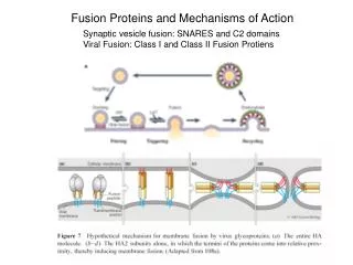
Class I and II Fusion Proteins. To reproduce, enveloped viruses must enter a host cell by fusing their own membrane coat with that of the cell. Fusion is caused by specific proteins in the viral membrane, and at least two classes of these proteins have been identified.

E N D
Class I and II Fusion Proteins • To reproduce, enveloped viruses must enter a host cell by fusing their own membrane coat with that of the cell. • Fusion is caused by specific proteins in the viral membrane, and at least two classes of these proteins have been identified. • In both classes, tightly regulated conformational changes are involved in membrane fusion.
Class I Fusion Proteins • Class I fusion proteins are composed of three identical protein subunits, the functional forms of which are generated from a precursor that is cleaved into two pieces. The carboxy-terminal end of one piece is anchored to the viral membrane; the other end (the new amino terminus) has a characteristic stretch of 20 hydrophobic amino acids — the fusion peptide. • An essential feature of class I proteins is the formation of a trimeric helical coiled-coil rod adjacent to the fusion peptide. This structure may act as a template for the refolding of protein segments near to the segments anchored in the virus membrane.
Class I Fusion Proteins • After binding to a receptor on the cellular membrane, or on exposure to the low pH found in intracellular compartments (endosomes), the protein forms an extended conformation and the hydrophobic fusion peptide inserts into the target membrane. • Several trimers are thought to be involved. • Protein refolding begins. The free energy thereby released causes the membranes to bend towards each other. • Formation of a restricted hemifusion stalk allows the lipids in the outer leaflets of the membranes to mix. • Protein refolding completes, forming the final, most stable form of the fusion protein,with the fusion peptide and transmembrane domain anti-parallel to each other but in the same membrane.
Class II Fusion Proteins • Class II fusion proteins have distinctly different structural features: they are predominantly non-helical, instead having a β-sheet-type structure; they are not cleaved during biosynthesis; and the portion that inserts into the target membrane is thought to be an internal hydrophobic fusion loop. • The proteins have a three-domain architecture, in which domain I begins at the amino terminus, domain II contains the internal fusion loop, and domain III is at the carboxy terminus
Class II: Dengue, Tick-Borne Encephalitis, Semliki Forest Viruses
Class II Fusion Proteins • The dimeric E protein binds to a cellular receptor and the virus is internalized to endosomes. • The acidic pH inside endosomes causes domain II to swing upward, permitting E monomers to rearrange laterally. • The fusion loop inserts into the outer leaflet of the host-cell membrane, enabling trimer formation. • The formation of trimer contacts extends from the top to the bottom of the molecule. Domain III shifts and rotates to create contacts, bending the membrane. • The formation of further contacts leads to unrestricted hemifusion and the final most stable form of the protein.