Bacteria
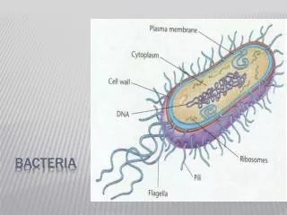
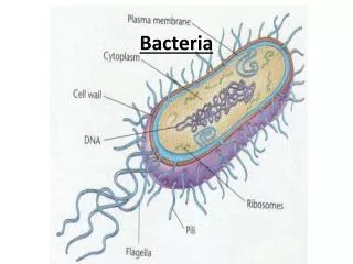
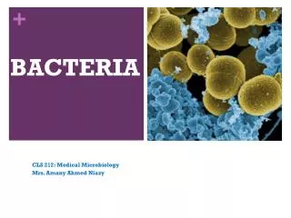
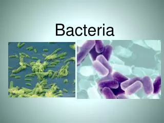
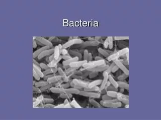
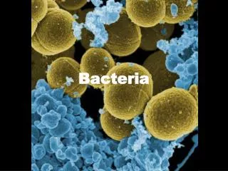
Bacteria. Staphylococcus bacteria in nose. Structure. Unicellular Some species form colonies 0.5–10 µ m, much smaller than the 10–100 µ m of many eukaryotic cells Most common shapes: spheres (cocci) rods (bacilli) and spirals. Bacterial Cell Wall. maintains cell shape

Bacteria
E N D
Presentation Transcript
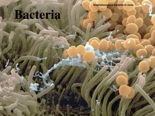
Bacteria Staphylococcus bacteria in nose
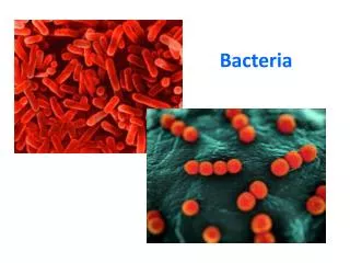
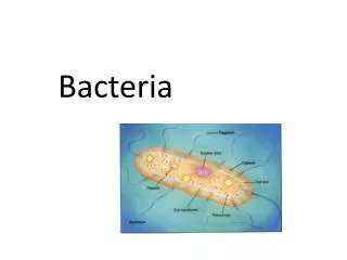
Structure • Unicellular • Some species form colonies • 0.5–10 µm, much smaller than the 10–100 µm of many eukaryotic cells • Most common shapes: • spheres (cocci) • rods (bacilli) • and spirals
Bacterial Cell Wall • maintains cell shape • provides physical protection, • prevents the cell from bursting in a hypotonic environment • Bacterial cell walls contain peptidoglycan, a network of sugar polymers cross-linked by polypeptides (proteins) sugar sugar polypeptide
Gram Stain Classification • Using the Gram stain, scientists classify bacterial into Gram + or Gram - groups based on cell wall composition • Gram - bacteria: • less peptidoglycan • outer membrane that can be toxic more likely to be antibiotic resistant • Many antibiotics target peptidoglycan and damage bacterial cell walls Gram - Gram +
Peptidoglycan layer Cell wall Plasma membrane Protein (a) Gram-positive: peptidoglycan traps crystal violet.
Carbohydrate portion of lipopolysaccharide Outer membrane Cell wall Peptidoglycan layer Plasma membrane Protein (b) Gram-negative: crystal violet is easily rinsed away, revealing red dye.
Gram- positive bacteria Gram- negative bacteria 20 µm
Structure • A polysaccharide or protein layer called a capsule covers many bacteria
Bacterial Motility • Most motile bacteria use flagellato propel themselves • Many exhibit taxis, the ability to move toward or away from certain stimuli Video: Prokaryotic Flagella (Salmonella typhimurium)
Flagellum Filament 50 nm Cell wall Hook Basal apparatus Plasma membrane
Fimbriae Fimbriae (also called attachment pili) • allows them to stick to their substrate or other individuals in a colony
Internal Organization • lack complex compartmentalization (ex. no nucleus, ER, mitochondrion, etc.) • Often perform metabolic functions using highly folded extensions of plasma membrane
1 µm 0.2 µm Respiratory membrane Thylakoid membranes (a) Aerobic prokaryote (b) Photosynthetic prokaryote
Genome • Most of the genome consists of a circular chromosome located in a nucleoid region • Some have smaller rings of DNA called plasmids • Plasmids w/ short codes of DNA that may be beneficial to bacteria (Ex. some code for antibiotic resistance)
Genome • Sex pili are longer than fimbriae and allow prokaryotes to exchange DNA
Plasmid F factor • Cells containing the F plasmid function as DNA donors during a process called conjugation • Cells without the F factor function as DNA recipients during conjugation • The F factor is transferable during conjugation
Plasmid F factor Bacterial chromosome F plasmid F+ cell Mating bridge F– cell Bacterial chromosome (a) Conjugation and transfer of an F plasmid
Plasmid F factor Bacterial chromosome F plasmid F+ cell Mating bridge F– cell Bacterial chromosome (a) Conjugation and transfer of an F plasmid
Plasmid F factor Bacterial chromosome F plasmid F+ cell F+ cell Mating bridge F– cell F+ cell Bacterial chromosome (a) Conjugation and transfer of an F plasmid
R Plasmids and Antibiotic Resistance • R plasmids carry genes for antibiotic resistance • Antibiotics select for bacteria with genes that are resistant to the antibiotics
Reproduction and Adaptation • Quick reproduction by binary fission • every 1–3 hours • Beneficial mutations can accumulate rapidly in a population, allowing for rapid evolution • ex. Antibiotic resistant strains are becoming more common • Many form inactive endospores • to remain viable in harsh conditions
Endospore 0.3 µm
Genetic Recombination • DNA from different individuals can be brought together by conjugation, transformation, transduction
Transformation and Transduction • A prokaryotic cell can take up and incorporate foreign DNA from the surrounding environment in a process called transformation • Transduction is the movement of genes between bacteria by vectors like bacteriophages (viruses that infect bacteria)
Phage DNA A+ B+ A+ B+ Donor cell
Phage DNA A+ B+ A+ B+ Donor cell A+
Phage DNA A+ B+ A+ B+ Donor cell A+ Recombination A+ A– B– Recipient cell
Phage DNA A+ B+ A+ B+ Donor cell A+ Recombination A+ A– B– Recipient cell A+ B– Recombinant cell
Pathogenic Prokaryotes • Prokaryotes cause about half of all human diseases • Lyme disease is an example
Fig. 27-21a Deer tick
Fig. 27-21c Lyme disease rash
Pathogenic Prokaryotes • Bacteria typically cause disease by releasing exotoxins or endotoxins • Exotoxins cause disease even if the prokaryotes that produce them are not present • Endotoxins are released only when bacteria die and their cell walls break down • Many pathogenic bacteria are potential weapons of bioterrorism