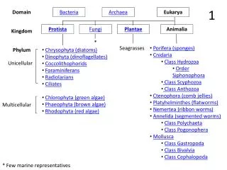
BACTERIA
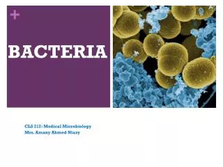
BACTERIA. CLS 212: Medical Microbiology Mrs. Amany Ahmed Niazy. Microorganisms. Cellular. Acellular. Prokaryotes. Eukaryotes. Viruses. Archaea. Fungi . . Bacteria. P rotozoae. Vir. Bacteria are the smallest and most versatile independently living cells known. . Prokaryotes.

BACTERIA
E N D
Presentation Transcript
BACTERIA CLS 212: Medical Microbiology Mrs. Amany Ahmed Niazy
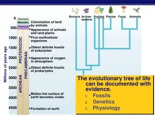
Microorganisms Cellular Acellular Prokaryotes Eukaryotes Viruses Archaea Fungi . Bacteria Protozoae Vir
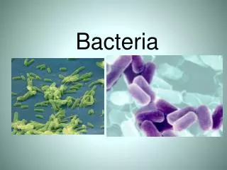
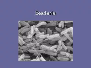
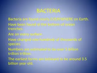
Bacteria are the smallest and most versatile independently living cells known.
Prokaryotes • Prokaryotic cells possess simpler structures than eukaryotic cells, since they do not have a nucleus or many cytoplasmic organelles. • There are two major types of prokaryotes: • Bacteria. • Archaea (also called archaebacteria) are often found in extreme environments (Oxygen-free environments concentrated salt-water hot, acidic water), and while they are clearly prokaryotic, they have evolved separately from bacteria.
History • Bacteria were first observed by Anton van Leeuwenhoek in 1676, using a single-lens microscope of his own design. • Robert Koch worked on cholera, anthrax and tuberculosis. • In his research into tuberculosis, Koch finally proved the germ theory, for which he was awarded a Nobel Prize in 1905.
Introduction • Bacteria (plural), Bacterium(singular). • Bacteriology The study of bacteria. • Bacteria are unicellular microscopic prokaryotes. • Bacteria are ubiquitous in every habitat on Earth, growing in soil, acidic hot springs, radioactive waste, water, and deep in the Earth's crust, as well as in organic matter and the live bodies of plants and animals. • Bacteria are vital in recycling nutrients such as the fixation of nitrogen from the atmosphere and decomposition of dead organic materials.
Classification of Bacteria • Bacteria can be classified on the basis of cell structure, cellular metabolism or on differences in cell components such as DNA, fatty acids, pigments, and antigens. • The most common method to classify pathogenic bacteria is on the basis of Gram Staining and Shape.
Classify pathogenic bacteria is on the basis of Gram Staining and Shape. Gram Stain Gram Positive Gram Negative Cocci Rods Cocci Rods
Taxonomy • The International Committee on Systematic Bacteriology (ICSB)maintains international rules for the naming of bacteria and taxonomic categories and for the ranking of them in the International Code of Nomenclature of Bacteria. Kingdom Bacteria PhylumProteobacteria Class Gamma Proteobacteria OrderEnterobacteriales FamilyEnterobacteriaceae Genus Escherichia Species Escherichia coli e.g. Escherichia coli
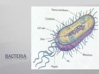
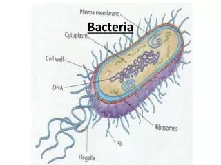
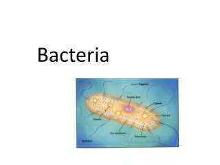
Exterior Structures • The exterior structure is made up of two to three layers: • In some species of bacteria an outer capsule. • Cell wall. • Cytoplasmic membrane.
Capsule • Some bacteria surround themselves with Capsule. • Most capsules are polysaccharides made of single or multiple types of sugar. • Capsule do not contribute to growth and multiplication. • Capsules provide some general protection for bacteria eg. Protect it from drying. . • Capsule major function in pathogenic bacteria is protection from the immune system. The capsule is a major virulence factor in the major disease-causing bacteria, such asStreptococcus pneumoniae. (Noncapsulated mutants of these organisms are avirulent, i.e. they don't cause disease).
Cell Wall – Why is it important? • The rigid cell wall gives the bacterium its shape and surrounds the cytoplasmic membrane, protecting it from the environment. • The strength of the wall is responsible for keeping the cell from bursting when there are large differences in osmotic pressure between the cytoplasm and the environment. • It also helps to anchor appendages like the pili and flagella, which originate in the cytoplasmic membrane and protrude through the wall to the outside.
Structure of Cell Wall The cell wall of bacteria is composed of peptidoglycan, which covers the entire surface of the cell. It is made up of a combination of peptide bonds and carbohydrates (protein-sugar) • Peptidoglycan is a huge polymer of interlocking chains of identical monomers. The backbone of the peptidoglycan molecule is composed of two derivatives of glucose: N-acetylglucosamine (NAG) and N-acetlymuramic acid (NAM). The NAG and NAM strands are connected by interpeptide bridges.
Structure of Cell Wall • The wall of a bacterium is classified in two ways: • Gram-positive. A gram-positive cell wall has many layers of peptidoglygan (up to 90% of the cell wall). Which makes it retain the crystal violet dye when the cell is stained. This gives the cell a purple color when seen under a microscope. • Gram-negative. The cell walls of gram negative bacteria are more chemically complex. Peptidoglycan makes up only 5 – 20% of the cell wall, and is not the outermost layer, but lies between the plasma membrane and an outer membrane. This outer membrane is similar to the plasma membrane, but is less permeable and composed of lipopolysaccharides (LPS). LPS is a harmful substance classified as an endotoxin.
Peptidoglycan and Antibiotics • Some antibiotics such as Penicillins and Cephalosporins, interfere with the linking of the interpeptides of peptidoglycan, but because of the LPS membrane, these antimicrobials can’t access the peptidoglycan of gram-negative bacteria. While gram-positive bacteria, are more susceptible to these antibiotics because of the lack of the LPS (lipopolysaccharides). Since the eukaryotic cells of humans do not have cell walls, our cells are not damaged by these drugs. Microorganisms that do not contain peptidoglycan.
Gram Positive Cell Wall • Contains two major components: • Peptidoglycan provide resistance to most human enzymes • Teichoic acids promote adhesion and anchor wall to membrane.
Gram Negative Cell Wall • The amount of peptidoglycan has been greatly reduced, it can be only one layer sheet. • Outer membrane (like all membrane) consist of phospholipids. It serves as a barrier to the passage of many molecules. Thus, it serves as protective barrier against certain antimicrobial medications.
GRAM STAIN • There are two main types of bacterial cell walls, Gram positive and Gram negative, which are differentiated by their Gram staining characteristics. • Gram stain Procedure: • Crystal violet • Iodine • Alcohol • Saffranine
Gram +veandGram –veCell Wall • The Gram positivecell wall is characterized by the presence of a very thickpeptidoglycan layer, which is responsible for the retention of the crystal violet dyes during the Gram staining procedure. Color of Bacteria:Blue-violet • The Gram negativecell wall contains a thinpeptidoglycan layer, which is responsible for the cell wall's inability to retain the crystal violet stain upon decolourisation with ethanol during Gram staining. Color of Bacteria:RED
Cell Membrane (=cytoplasmic membrane) Cell membrane is composed of phospholipids bilayer and proteins which is found through out the living world. • It is the site of synthesis of DNA and other material. • It is responsible for selective and active transport of materials in and out of the cell. • It is involved in secretion of some exotoxins and hydrolytic enzymes involved in the pathogenesis of disease.
Flagella, Appendages • Flagella (singular, flagellum) are long hair like protein structure that are found in many species of bacteria. • They may be found at either or both ends of a bacterium or all over its surface. • Function of the Flagella: the flagella beat in a propeller-like motion to help the bacterium move toward nutrients, or away from toxic chemicals.
Pili, Appendages • Pili(singular, pilus) are short hair-like projections found all around the surface of cells of many bacteria. • Function of the Pili: the pili assist the bacteria in attaching to other cells and surfaces, such as teeth, intestines, and rocks. • Without pili, many disease-causing bacteria lose their ability to infect because they are unable to attach to host tissue.
Cytosol (cytoplasm of the bacteria) • It is where the functions for cell growth, metabolism, and replication are carried out. • It is a gel-like matrix composed of water, enzymes, nutrients, wastes, and gases and contains cell structures such as ribosomes, a Nucleoid, and plasmids. • The cell envelope encases the cytoplasm and all its components. • Unlike the eukaryotic (true) cells, bacteria do not have a membrane enclosed nucleus.The chromosome, a single, continuous strand of DNA, is localized, but not contained, in a region of the cell called the nucleoid. All the other cellular components are scattered throughout the cytoplasm.
The Nucleoid The nucleoid is a region of cytoplasm where the chromosomal DNA (chromosome) is located. • The chromosome of prokaryotes is an irregular mass within the cytoplasm, that is usually attached to the cytoplasmic membrane. • It is a large, circular molecule of double-stranded DNA. • The absence of nuclear membrane is very important for rapid growth or prokaryotic cells in changing environments. Nucleoid
Ribosomes Ribosomes Involved in protein synthesis, they translate the genetic code form nucleic acid to that of amino acids • It is much more abundant than in the cytoplasm of eukaryotic cells this is a reflection of the higher growth rate of bacteria. • Ribosomes of prokaryotic cells (30S – 50S) are smaller in size than ribosomes of eukaryotic cells (80S). • They differ in structure wich make them a target for certain antibiotics.
Plasmids • Plasmids are small usually circular, double-stranded DNA. • It is separated from the chromosome, and they are not involved in reproduction. • They are found in many strains of bacteria. • A single bacterial cell can harbor multiple types of plasmids. Plasmids replicate independently of the chromosome and, while not essential for survival, appear to give bacteria a selective advantage. For example, many plasmids code for the production of one or more enzymes that destroy certain antibiotics (resistance to that antibiotic).
How are plasmids passed on from one bacteria to the other?? • Plasmids are passed-on to other bacteria through Two ways: 1. For most plasmid types, copies in the cytoplasm are passed on to daughter cells during binary fission.
How are plasmids passed on from one bacteria to the other?? • Other types of plasmids form a tube-like structure at the surface called a pilus that passes copies of the plasmid to other bacteria during conjugation, a process by which bacteria exchange genetic information.
Plasmids • Many plasmid genes promote survival and pathogenesis. • Plasmid are responsible for transfer of cellular properties such as, production of toxins, production of pili , resistance to antimicrobials and other toxic chemicals. • The ability to insert specific genes into plasmids have made them extremely useful tools in the fields of molecular biology and genetics, specifically in the area of genetic engineering.
Endospores • Endospores are bacterial survival structuresthat are highly resistantto many different types of chemical and environmental stresses and therefore enable the survival of bacteria in environments that would be lethal for these cells in their normal vegetative form. • Resistance of spore is due to dehydrated state, and specialized coats. • Germination of spores reproduces cell identical to that which was sporulated.
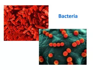
Cell Morphology & Shape of Bacteria Coccus(spherical):e.g. Streptococci, Staphylococci Spirillum(spiral): e.g.Treponemaspp. Bacillus (rod-like): e.g.Enterobacteriaceaspp.
Bacteria typically have one of three shapes: • spheres (cocci). • rods (bacilli), • spiral (spirilla). • Unicellular, they often stick together forming clumps or filaments.
Replication of Bacteria • Bacterial cells replicate asexuallyby a process called: Binary Fission. • One cell doubles in size and splits in half to produce two identical daughter cells. These daughter cells can then double in size again to produce four sibling cells and these to produce eight, and so on. • Doubling Time: the time it takes for a bacterial cell to grow and divide in two. • When nutrients are plentiful, the doubling time of some bacterial species can be as short as 20 minutes. However, most bacterial species show a doubling time between 1-4 hours.
Replication DNA Duplicate of original molecule Transcription Replication of Chromosomal DNA of Prokaryotes RNA Translation Protein
Replication of Bacteria • The cytoplasm of a bacterial cell contains the DNA moleculesthat make up the bacterial genome. • Transcriptional machinerycopies DNA into ribonucleic acid (RNA). • Ribosomestranslate the messenger RNA information into proteinsequence.