Download
1 / 16
300 likes | 1.19k Vues
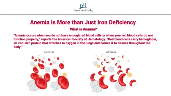
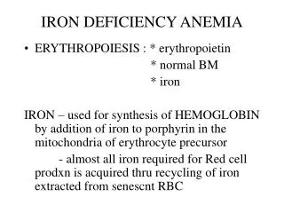
IRON DEFICIENCY ANEMIA. ERYTHROPOIESIS : * erythropoietin * normal BM * iron IRON – used for synthesis of HEMOGLOBIN by addition of iron to porphyrin in the mitochondria of erythrocyte precursor

E N D
IRON DEFICIENCY ANEMIA • ERYTHROPOIESIS : * erythropoietin * normal BM * iron IRON – used for synthesis of HEMOGLOBIN by addition of iron to porphyrin in the mitochondria of erythrocyte precursor - almost all iron required for Red cell prodxn is acquired thru recycling of iron extracted from senescnt RBC
IDA • Maximal Iron Absorption – DUODENUM & UPPER JEJUNUM – where acidic gastric juices reduces insoluble FERRIC IRON to its soluble FERROUS state • Iron Absorption Regulation – Cytoplasmic iron concentrate in mucosal cells
IDA • Measurement of Iron Supply: 1. Serum Iron- amount of iron bound to transferrin 2. TIBC- measure of total binding capacity of transferrin. Decrease iron = inc TIBC 3. Serum ferritin – Iron + apoferritin – protein which binds to free ferrous iron 4. Marrow iron stores
IDA • IRON Transport: 1. Pinocytosis 2. Transferrin – principal means of moving iron IRON Storage: 1. Ferritin – enclosed in a shell composed of a protein (apoferritin) 2. Hemosiderin – aggregates of ferritin molecules thar have been stripped of apoferritin
INCREASE Acids HCl Vit C Inorganic Iron Ferrous Iron Iron deficiency Increased demand Primary Hemochromatosis DECREASE Alkalis Antacids Pancreatic secretions Organic Iron Ferric Iron Excess Iron Decreased Utilization Infection/Inflamation Factors that Influence Iron Absorption
Major Causes of IDA 1. Chronic blood loss – most common • 2. Increased Iron Requirement • 3. Iron Malabsorption • 4. Inadequate dietary iron intake
Stages of IDA • I. Storage Iron Depletion iron reserves are lost w/o compromise of the iron supply for erythropoiesis BM aspirate- decrease/absent iron stain decrease level of serun ferritin II. Iron Deficient Erythropoiesis erythroid iron supply is reduced w/o development of anemia RBC- microcytic, hypochromic increase TIBC III. Iron Deficiency Anemia severe hypochromic and microcytic RBC
Clinical Mx of IDA • Non specific – fatigue, weakness • Signs – pallor, tachycardia, unexplained retinal hemorrhages and splenomegaly • Atrophic changes in epithelium: a. oral lesion, angular cheilosis, glossitis, stomatitis b. dysphagia c. koilonychia d. pica
Parenteral Iron Therapy • 1. Cannot tolerate the side effects of oral therapy • 2. Suffers from inflammatory bowel dss/peptic ulcer • 3. Does not comply with prescribed dosages • 4. Displays documented iron malabsorption • 5. Suffer from a condition such as hereditary hemorrhagic telangiectasia
Management of IDA • FeSo4 – 50 mg elemental iron/ 325 mg tab • Ferrous gluconate/fumarate – better tolerated Reticulocytosis – 3-4 days after initiation of iron therapy
Replacement Therapy • No Response: 1. Incorrect diagnosis 2. Continued loss of iron 3. Chronic infection/inflammation 4. Lack of patient compliance 5. Ineffective release of iron 6. Malabsorption of iron
ANEMIA OF CHRONIC DISEASE • Mechanism of Action 1. Relative Iron Deficiency: a. Apolactoferrin – iron binding protein released into the bloodstream by phagocytes in response to inflammation, strips iron from transferrin and return it to mononuclear phagocytes w/c reconverts iron to ferritin and hemosiderin
ANEMIA OF CHRONIC DSS b. Interleuken-1 – released by monocytes and macrophages. Stimulates increased retention of iron by macrophages to limit amount of iron for bacterial growth, limiting also iron for erythropoiesis 2. Shortened life span – 60-90 days 3. Relative marrow failure – low EPO levels/ ability of erythroid precursor to respond to EPO is impaired (BFU-E)
ANEMIA OF CHRONIC DSS • Etiology : 1-2 months of sustained dss 1. Chronic ifection/inflammatory dss.: TB, Pneumonia, osteomyelitis, bacterial endocarditis 2. Chronic non infectious inflammatory dss: sarcoidosis,SLE, RA 3. Malignancies – CA, lymphoma, sarcoma
Anemia of Chronic Dss Clinical Mx.: normocytic, normochromic Hct- 25-35% ; normal WBC/PC usually asymptomatic DIAGNOSIS: 1. Chronic inflammatory dss/malignancy 2. Low/normal level of serum iron asso with decrease TIBC and transferrin saturation 3. Normal/incrse serum ferritin 4. Abundant hemosiderin
Anemia of Chronic Dss • Treatment: * Treat underlying cause * EPO therapy : 100-150 u/kg EPO TIW, SC or IV * Iron is contraindicated