Postpartum Hemorrhage
Postpartum Hemorrhage. Learning Objectives. Define and differentiate between early and late PPH. Identify causes and risk factors of PPH. Identify symptoms and signs of PPH and list the laboratory investigations.

Postpartum Hemorrhage
E N D
Presentation Transcript
Learning Objectives • Define and differentiate between early and late PPH. • Identify causes and risk factors of PPH. • Identify symptoms and signs of PPH and list the laboratory investigations. • Manage PPH immediately after delivery of the baby and before delivery of the placenta. • Manage PPH immediately after delivery of the placenta. • Manage PPH 24 hours after delivery.
Problem in Egypt • Postpartum hemorrhage is the single greatest cause of maternal death in Egypt. • Postpartum hemorrhage was responsible for 34% of all maternal deaths. • The most important avoidable factors were: • Substandard care by obstetricians(50%) • Lack of blood (31%) • Substandard care by dayas (14%)
Definition • Postpartum hemorrhage is excessive blood loss after delivery sufficient to affect the general condition of the mother as shown by tachycardia and/or hypotension. • The traditional definition, based on a blood loss 500 mL from or within the reproductive tract after delivery, is difficult to estimate in clinical practice.
Average Blood Loss and Complexity of Delivery • Vaginal delivery–500 ml • Cesarean section–1000 ml • Repeat cesarean section & TAH–1500 ml • Emergency hysterectomy–3500 ml. Pritchard AJOB 1961 Clark Obstet Gynecol 1984
Types of PPH • Primary: • within the first 24 hours • Secondary: • after the first 24 hours up to the 42nd day
Causes of PPH • Uterine atony • Genital tract trauma • Third stage complications: • Mismanagement of the third stage of labor • Acute inversion of the uterus • Abnormal or incomplete placental separation • Coagulation disorders
Factors Predisposing To Uterine Atony • Overdistended uterus • Uterine muscle exhaustion • Amniotic infection • Functional or anatomic distortion of the uterus due to a fibroid uterus, placenta previa or uterine anomalies • Certain general anesthetics (e.g. halothane) • History of previous PPH
Factors Predisposing To Genital Tract Trauma • Precipitate delivery • Operative or manipulative delivery • Malposition or deep engagement of the fetal head predisposes the genital tract to extensions and/or lacerations during a CS • Previous uterine surgery predisposes to a uterine rupture
Factors predisposing to retained products of conception • An incomplete placenta at delivery • Previous uterine surgery (morbid adhesions) • High parity • An abnormal placenta on ultrasound • Placenta previa • Placenta implanted on a scar • Placenta increta,percreta, accreta • Accessory placental lobe • Placental Abruption
Abnormalities of Coagulation • Pre-existing coagulation disorders such as: • Hemophilia A or Von Willebrand’s disease • Idiopathic thrombocytopenic purpura (ITP) • History of liver disease • Use of anticoagulants • Acquired in pregnancy: • Thrombocytopenia with pre-eclampsia (HELLP syndrome) • DIC caused by: • Abruptio placentae • Chorioamnionitis • IUFD
Diagnosis In a woman with excessive PPH, begin first aid management while simultaneously taking a history and performing a physical examination. • History. • General examination: • Tachycardia and hypotension • Abdominal examination • Consistency of the uterus (lax or firm) • Uterine fundal level. • Any abdominal tenderness or rigidity • Scars from previous operations
Diagnosis - (Cont.) • Local examination: • Assess the amount of vaginal bleeding • Look for lacerations (perineal, vulvar, vaginal or cervical) • Determine whether the placenta has been delivered • Tachycardia and hypotension may be present without evidence of excessive blood loss in cases of uterine rupture. • In a patient with hypertension or pre-eclampsia, severe blood loss may cause a misleading “normal” blood pressure reading.
Laboratory Investigations • ABO grouping and Rh type • Cross-match at least two units of whole blood or as needed for the clinical situation. • CBC (Hb, Hct, differential, platelet count) • Bleeding time • Coagulation time
Prophylactic Measures • During the antenatal period: • Recognize risk factors • Diagnose anemia at an early stage • Ensure iron and folic acid supplementation • Educate the patient regarding the following: • Anemia prevention • Going to the hospital early in labor • Seeking immediate medical attention if vaginal bleeding occurs
Prophylactic Measures - (Cont.) • During labor and delivery: • Identify patients at risk of developing PPH • If the hemoglobin level is 8 gm/dL, cross-match at least one unit of whole blood even if the delivery is normal, or two units if it is CS, to be used if needed. • Actively manage the third stage of labor. • Administer 10 IU of oxytocin IM with the delivery of the anterior shoulder. • Perform controlled cord traction to deliver the placenta. • Perform uterine massage.
First Aid Management • SHOUT FOR HELP • Insert two wide bore IV cannulae (size 16 or 18), and withdraw 20 mL of blood for cross-matching and required investigations before starting any IV fluids • IV crystalloid solution at a fast drip (1 L/hour) and start ecbolic drugs • Continuously monitor pulse and BP every five minutes. • Insert a Foley catheter. • Monitor urine output. • Massage the uterine fundus.
Ecbolic Drugs • Misoprostol: • Rectal 1000 g tablets [200mcgX5] • Oxytocin: • IV Infusion 20 IU in 1 L IV fluids at 60 drops per minute then infuse at 40 drops per minute • Not more than 3 L of IV fluids containing oxytocin • Do not give as an IV bolus • Methyl ergometrine: • IM or IV (slowly): 0.2 mg, repeat after 15 minutes. • If required, give 0.2 mg IM or IV (slowly) every 4 hours, maximum 5 doses (Total 1.0 mg) • Contraindications: pre-eclampsia, hypertension, heart disease
Active Management • Hemorrhage after delivery of the neonate, before delivery of the placenta (retained placenta): • Determine the cause of the retained placenta (hourglass contraction versus partial separation). • Administer Ergotrate 0.2 mg (1 ampule) IM , or give misoprostol 200 µg (one tablet) rectally, followed by controlled cord traction to deliver the placenta.
Active Management – (Cont.) • Hemorrhage after delivery of the neonate, before delivery of the placenta (retained placenta): • Avoid forceful cord traction and fundal pressure as they may cause uterine inversion. • Explore the perineum, vagina, and cervix, looking for lacerations if the bleeding persists without delivery of the placenta. • Call the anesthesiologist. • Manually remove the placenta with adequate sedation (e.g., Valium or Pethidine) or anesthesia.
Manual Removal of the Placenta Introducing one hand into the vagina along the cord
Manual Removal of the Placenta - (Cont.) Supporting the fundus while detaching the placenta
Manual Removal of the Placenta - (Cont.) Withdrawing the hand from the uterus
Manual Removal of the Placenta - (Cont.) • Palpate the inside of the uterine cavity to ensure that all placental tissue has been removed • Give ecbolic drugs • Massage the fundus of the uterus • Examine the uterine surface of the placenta to ensure that it is complete • Examine the woman carefully and repair any tears or episiotomy. • If the placenta does not separate from the uterine surface, suspect an adherent placenta and proceed to perform a laparotomy and possibly a subtotal hysterectomy.
Hemorrhage Immediately After Delivery of Placenta • First Aid Management • Ecbolics • EUA if bleeding persists • Explore the perineum, vagina, cervix and uterus looking for lacerations and repair as needed • Explore the uterine cavity for retained placental fragments • If there are retained placental fragments, remove them manually or with ring forceps • If the cervical tear has extended deep beyond the vaginal vault (incomplete uterine rupture), a laparotomy may be required.
Hemorrhage Immediately After Delivery of Placenta • If the hemorrhage still persists, assess the patient’s clotting status using a bedside clotting test. • Failure of a clot to form after 7 minutes or a soft clot that breaks down easily suggests coagulopathy. • If bleeding continues in spite of the above management, perform bimanual compression of the uterus. • Perform an emergency laparotomy if the above maneuver fails or if a ruptured uterus is found. Do not waste valuable time trying to save the uterus at the expense of the general condition of the mother.
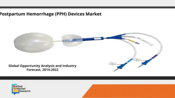
Uterine Packing (Balloon Tamponade) • Using either a Foley catheter or a Sengstaken-Blakemore tube • Useful for uterine atony, retained placental tissue, and placenta accreta • Both the Foley catheter and the Sengstaken-Blakemore tube have open tips, which permit continuous drainage from the uterus
Uterine Packing (Balloon Tamponade) • Ultrasound can more effectively detect a developing hematoma when the contrast is a fluid-filled balloon as opposed to blood-saturated gauze. • Thus, this technique has the advantage of being not only therapeutic but also diagnostic when used in combination with ultrasound, in differentiating the various etiologies described above.
Uterine Packing (Balloon Tamponade) • Foley catheter procedure: • Using a 24 Fr Foley catheter, guide the tip into the uterine cavity and inflate with 20 to 30 mL of saline. • Sengstaken-Blakemore tube: • Has an advantage due to the larger capacity of its balloon tip • If the bleeding stops, the patient can be observed with the catheters in place and then they are removed after 12 to 24 hours.
Laparotomy • Inspect the uterus. If lacerated, repair adequately and if it is atonic, perform direct uterine massage • Surgical compression suture (B-Lynch suture) technique • Uterine packing (balloon tamponade) • Ligation of the uterine and utero-ovarian arteries • Bilateral internal iliac artery ligation • Supravaginal hysterectomy
Surgical Compression Suture (B-Lynch suture) Technique • Mechanical compression of the uterine vascular sinuses prevents further engorgement with blood and continued hemorrhage • Used to treat atony and hemorrhage that does not respond to pharmacologic interventions • Used if bimanual compression decreases the amount of uterine bleeding by abdominal and perineal inspection • Although originally described using No. 2-0 chromic catgut, variations using No. 0 Vicryl suture have been equally successful
Surgical Compression Suture (B-Lynch suture) Technique - (Cont.)
Ligation of Uterine , Utero-Ovarian and Hypogastric Arteries • The obstetrician should consider ligation of the hypogastric arteries if trained in this technique
Secondary PPH (severe degree) • First aid management • Administer IV broad spectrum antibiotics • Give ecbolic drugs • If the cervix is dilated, explore by hand to remove large clots and placental fragments, plus manual exploration of the uterus • If the cervix is not dilated, evacuate the uterus to remove placental fragments • In rare cases, if bleeding continues, consider performing a uterine and utero-ovarian artery ligation or a hysterectomy
Secondary PPH (mild degree) • Check the cervix: • If the cervix is open, perform curettage and perform a histological investigation of the products of the curetting. • If the cervix is closed give broad spectrum antibiotics for one week and reassess the condition. • Perform a histological investigation of the products of the curetting or hysterectomy specimen, if possible, to rule out a gestational trophoblastic tumor.
Monitoring During Hospital Stay • Check the patient’s blood pressure and pulse every 30 minutes for the first two hours, then hourly for six hours, and then every four hours. • Perform gentle uterine massage every 30 minutes. • Check for vaginal bleeding every hour. • Check urine output every two hours.
Remember that a postpartum patient can lose a large amount of blood in a very short time. You must act promptly and anticipate complications. • Assure adequate team coverage. • A laparotomy for PPH is an extremely urgent situation and need not be delayed while waiting for a blood transfusion. • Administer prophylactic antibiotics before and after the procedure. • Do not give oxytocin as an undiluted IV push since the patient may collapse.