MIC159 Microbial World
MIC159 Microbial World. Siti Sarah Jumali Room 3/14. Microorganism & Microbiology. Microorganism. Microbiology. Study of microorganisms Foundation of modern biotechnology Among the many specialized fields of microbiology

MIC159 Microbial World
E N D
Presentation Transcript
MIC159 Microbial World Siti Sarah Jumali Room 3/14
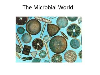
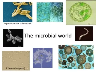
Microorganism & Microbiology Microorganism Microbiology Study of microorganisms Foundation of modern biotechnology Among the many specialized fields of microbiology -Virology, Mycology, Bacteriology, Immunology, Microbial Ecology, Biotechnological Microbiology, Environmental Microbiology, Food Microbiology, Forensic Microbiology, Molecular Biology • Living things which individually are too small to be seen with the naked eye. • All of the following may be considered microorganisms: • bacteria (eubacteria, archaebacteria) • fungi (yeasts, molds) • protozoa • microscopic algae • viruses • various parasitic worms
Microorganism & Microbiology cont’d Two main themes involved in Microbiology 1- Basic- cellular processes 2-Applied- concerning agriculture, industry and health
Themes in Microbiology and its field Bacteriology Phycology Mycology Virology Parasitology Protozoalogy Microbial metabolism Microbial genetics Microbial genetics Immunology Epidemiology Etiology Environmental microbiology Food & beverage tech Pharmaceutical microbiology Genetic Engineering Infection control Chemotherapy
Microorganism • Too small • Germ-rapidly growing cell • Has habitat • Live in population (not alone) • Communities are either swimming freely or attached to a surface (biofilm) • Interact between communities; may either be - harmful (because of waste product) - beneficial (cooperative feeding efforts-wastenutrient)
Microbes in our lives • Some are pathogenic (disease-causing) • Decompose organic waste • Produces through photosynthesis (e.g.Purplesulphur bacteria must fix CO2 to live) • Play role in industry (e.g. fermentation to produce ethanol and acetone) • Produce fermented food (vinegar, cheese & bread) • Produce products used in manufacturing (cellulase) and treatment (insulin)
Microbes and agriculture • Nitrogen fixation • Rumen microbes help digest grass and hay in cows, sheep etc • Cycles nutrients (C, N and S) • Causes disease to animals and plants
Microorganism and Food • Microorganism and food 1) Prevent spoilage (tempeh, salted fish) 2) Assist in manufacturing of food • Microorganisms and energy 1) Natural gas (methane) 2) Ethanol (biofuel) 3) Bioremediation • Microbes and the future 1)Genetic engineering
Thoughts for the day So, how can microbes benefit us? • In food? • In environment? • In preventing disease? • In agriculture? • In energy? • In waste-water treatment?
Naming and Classifying microorganisms Linnaeus system for scientific nomenclature Each organism has two names: 1) Genus 2) Specific epithet
Scientific Names • Italicized or underlined. The genus is capitalized, and the specific epithet is with lowercase • Could be as an honor for the scientist • A Latin origin e.g. Escherichia coli (E. coli) - discoverer: Theodor Escherich - describes the habitat (colon/intestine) e.g. Staphylococcus aureus(S. aureus) - Clustered (staphylo), spherical (cocci) - Gold colored colonies (aureus) In intestine On skin
Classification of bacteria Plants, animals, Fungi, Protists Microorganisms Also include fungi, protozoa, algae, viruses, multicellular animal parasites
Bacteria (P)/ Bacterium (S) Archaea Prokayotic Lack peptidoglycan Live in extreme environments Include - Methanogens - Extreme halophiles - Extreme thermophiles • Prokaryotes • Has peptidoglycan cell walls • Binary fission • Utilize organic/inorganic chemicals, or photosynthesis to obtain energy
Types of Eukaryotes Protozoa Algae Unicellular/multicellular eukaryote Has cellulose cell walls Gain energy through photosynthesis Produce molecular and organic compounds • Unicellular eukaryote • Absorb or ingest organic chemicals • May move using pseudopods, cilia or flagella • e.g. Amoeba
Fungi (singular: Fungus) • Eukaryotes • Chitin cell walls • Use organic chemicals for energy • Molds and mushrooms are multicellular, consists of mycelia (composed of filaments called hyphae) • Yeasts are unicellular
Viruses Multicellular animal parasites Helminths: flatworms and roundworms Multicellular • Too small to be observed with light microscope • Consists of DNA/RNA core • Core is surrounded by protein coat • Coat may be enclosed in a lipid envelope • Viruses are replicated only when they are in living host cell • Bacteriophage-viruses that infect bacteria • Viroids-nucleic acid without protein coating • Prions- Infectious protenacious particles
How do we view microorganisms? • Units of measurement When talking about cells and microscopic organisms, you would be measuring using MICROMETRE (abbreviated: µ --micron ) or stated as: µm (micrometer). 1 µm = 1 x 10⁻⁶ meters/ 1 x 10⁻³ mm 1 mm= 1 x 10³ nanometers/ 1 x 103 µm To give you the idea of how small a micro metre is, 1- a human hair is about 100 µm, wide, 2- a red blood cell would be around 8 µm wide 3- typical size of an animal cell would be from 10 - 100 µm
Microscope • Light microscope • Uses light • Few types • Compound light microscopy • Darkfield microscopy • Phase-contrast microscopy • Differential interference contrast microscopy • Fluorescence microscopy • Confocal microscopy
Compound light microscope • The image is magnified again by ocular lens Total magnification= objective lens x ocular lens • Resolution- ability of lenses to distinguish two points • e.g. RP of 0.4 nm can distinguish between 2 points ≥ 0.4 nm • Shorter light wavelength provides greater resolution • Refractive index- Light bending ability of a medium • Light may bend in air sthat it misses the small high-magnification lens • Immersion oil is used to keep the air from bending.
Types of Microscopes Light Microscope - found in most schools, use compound lenses and light to magnify objects. The lenses bend or refract the light, which makes the object beneath them appear closer. Stereoscope - this microscope allows for binocular (two eyes) viewing of larger specimens. (The spinning microscope at the top of this page is a stereoscope) Scanning Electron Microscope - allow scientists to view a universe too small to be seen with a light microscope. SEMs do not use light waves; they use electrons (negatively charged electrical particles) to magnify objects up to two million times. Transmission Electron Microscope - also uses electrons, but instead of scanning the surface (as with SEM's) electrons are passed through very thin specimens. Specimens may be stained with heavy metal salts
Magnification Your microscope has 3 magnifications: Scanning, Low and High. Each objective will have written the magnification. In addition to this, the ocular lens (eyepiece) has a magnification. The total magnification is the ocular x objective. Total magnification = magnification of eyepiece x magnification of objective lens
A Brief History of Microbiology Development of microscopy
Van Leeuwenhoek’s description of Bacteria From his teeth, he observed & (B)- rod forms (C) & (D)- motion pathway (E)- Spherical form (F)- Longer type of spherical form (H)- Cluster -Royal Society letter (Sept 17th, 1683) The microscope used Simple microscope (one lens)
Spontaneousgenerationcontroversy 1688: Francesco Redi(1626-1678) was an Italian physician who refuted the idea of spontaneous generation by showing that rotting meat carefully kept from flies will not spontaneously produce maggots. 1836: Theodor Schwann (1810-1882) helped develop the cell theory of living organisms, namely that that all living organisms are composed of one or more cells and that the cell is the basic functional unit of living organisms. 1861: Louis Pasteur's (1822-1895) famous experiments with swan-necked flasks finally proved that microorganisms do not arise by spontaneous generation.
The Golden Age of Microbiology ~1857-1914 (about 50 years) Beginning with Pasteur’s work, discoveries included relationship between microbes and disease, immunity, and antimicrobial drugs Robert Koch a. Identified a bacterium as cause of anthrax b. Introduced agar, inoculating loop to transfer bacteria and prepare pure cultures. c. Introduced “Koch’s Postulates” and the concept that a disease is caused by a single organism. Joseph Lister (1865) a. Introduced the “antiseptic technique”. b. Use of phenol (carbolic acid) as disinfectant. MartinusBeijerinck (1884 - 85) a. Discovered “viruses” (toxins, poisons). b. Infectious agents in tobacco plant fluids. Paul Ehrlich (1910) a. Introduced concept of chemotherapy. b. Use of salvarsan for the treatment of syphilis. Alexander Fleming (1928) a. Discovered the first antibiotic - penicillin.
Louis Pasteur's (1822-1895) famous experiments with swan-necked flasks • This eventually led to: • Development of sterilization • Development of aseptic technique
Proof that microbes cause disease 1546: Hieronymus Fracastorius(GirolamoFracastoro) wrote "On Contagion", the 1st known discussion of the phenomenon of contagious infection. 1835: AgostinoBassi de Lodi showed that a disease affecting silkworms was caused by a fungus - the first microorganism to be recognized as a contagious agent of animal disease. 1847: Ignaz Semmelweiss (1818-1865), a Hungarian physician-decided that doctors in Vienna hospitals were spreading childbed fever while delivering babies. He started forcing doctors under his supervision to wash their hands before touching patients. 1857: Louis Pasteur proposed the “Germ theory of disease”. • - Ancients believed that disease was the result of a divine punishment. Pasteur fought to convince surgeons that germs existed and carried diseases, and dirty instruments and hands spread germs and therefore disease. Pasteur's pasteurization process killed germs and prevented the spread of disease. 1867: Joseph Lister (1827-1912) introduced antiseptics in surgery. By spraying carbolic acid on surgical instruments, wounds and dressings, he reduced surgical mortality due to bacterial infection considerably. 1876: Robert Koch (1843-1910). German bacteriologist was the first to cultivate anthrax bacteria outside the body using blood serum at body temperature.
Robert Koch demonstrated the first direct role of a bacterium in disease Koch's postulates • "Koch's postulates" (1884), the critical test for the involvement of a microorganism in a disease: • The agent must be present in every case of the disease. • The agent must be isolated and cultured in vitro. • The disease must be reproduced when a pure culture of the agent is inoculated into a susceptible host. • The agent must be recoverable from the experimentally-infected host. • This eventually led to: • Development of pure culture techniques • Stains, agar, culture media, petri dishes
Preparing smears for staining • Staining- coloring microbe with a dye to emphasize certain structure • Smear- A thin film of a microbe solution on a slide, a smear is usually fixed to attach microbes to the slide and kill microbes
Staining • Stain usually consists of +ve and –ve ion • Basic dye- chromophore is a cation • Acidic dye- chromophore is an anion • Staining the background instead of the cell is called negative staining
Staining Simple stain Differential stain Distinguish Gram stain Acid-fast stain - staining with one dye - mordant may be used to hold the stain or to coat the specimen to enlarge it Gram stain Acid-fast stain Stained waxy cell wall is not decolorized by acid-alcohol • Mycobacterium • Nocardia Distinguish • Gram +ve and gram -ve • Gram +ve bacteria are prone to penicillin and detergents • Gram –ve are more resistant to antibiotics
Special stain Gram stain Distinguish • Gram +ve and gram -ve • Gram +ve bacteria are prone to penicillin and detergents • Gram –ve are more resistant to antibiotics Distinguish special parts of cells • Capsule • Endospore (Malachite green and safranin) • Flagella (carbolfuchsin simple stain) Gram –ve (pink)
So, microorganisms are something studied using characteristic techniques including: • aseptic technique • pure culture technique • microscopic observation of whole organisms Questions? Recommended reading: Microbiologyby L.M.Prescott et al. 6th edition.