Download
1 / 20
200 likes | 288 Vues
Learn about the flexors and extensors of the forearm, their origins, innervation, movements, and compartments. Explore the fascial sheath and muscles responsible for pronation and supination. Study the anatomy of the forearm in detail.

E N D
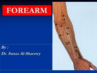
FOREARM By : Dr. Sanaa Al-Sharawy
OBJECTIVES At the end of this lecture, the student should able to : List the names of the Flexors Group of Forearm (superficial & deep muscles). Identify the common flexor origin of flexor muscles and their innervation & movements. Identify supination & poronationand list the muscles produced these 2 movements. List the names of the Extensor Group of Forearm (superficial & deep muscles). Identify the common extensor origin of extensor musles and their innervation & movements.
The forearm extends from elbow to wrist. • It posses two bonesradius laterally & Ulna medially. • The two bones are connected together by the interosseous membrane. • This membrane allows movement of Pronation and Supination while the two bones are connected together. • Also it gives origin for the deep muscles.
The forearm is enclosed in a sheath of deep fascia, which is attached to the posterior border of the ulna . • This fascial sheath, together with the interosseous membrane & fibrous intermuscular septa, divides the forearm into several compartments, each having its own muscles, nerves, and blood supply. Fascial Compartments of the Forearm
FLEXOR GROUP • These muscles: 8 • Act on the elbow & wrist joints and those of the fingers. • Form fleshy masses in the proximal part and become tendinous in the distal part of the forearm. • Arranged in three groups: I-Superficial: 4 • Pronator teres • Flexor carpi radialis • Palmaris longus • Flexor carpi ulnaris II-Intermediate: 1 • Flexor digitorum superficialis III- Deep: 3 • Flexor digitorum profundus • Flexor pollicis longus • Pronator quadratus
Superficial Flexors: • They arise - more or less- from the common flexor origin(front ofmedial epicondyle). • All are supplied by median nerve except one, flexor carpiulnaris, FCU (ulnar). • All cross the wrist joint except one, pronatorteres, (PT).
Flexor Carpi Radialis • Insertion:Base of 2nd metacarpal bone • Action: Flexion & abduction of the hand • Palmaris Longus • Insertion:into the flexor retinaculum & palmaraponeurosis. • Action: Flexes hand & tightens palmer aponeurosis • Pronator teres Insertion: middle of lat. surface of radius • Action: pronation & flexion of forearm .
Flexor Digitorum Superficialis • Origin: • Common flexor origin, • Coronoid process of ulna; • Anterior surface of radius • Insertion: • base of middle phalanges of medial 4 fingers. • Action: • Flexes middle and proximal phalanges of medial 4 fingers, and the hand • Flexor Carpi Ulnaris • Insertion: • Pisiform, • hook of hamate • 5th metacarpal bone • Action: • Flexion and adduction of the hand.
Deep Flexors • One above ulna: Flexor Digitorumprofundus • One above radius: Flexor pollicis longus • One above the 2 bones: Pronator Quadratus.
Pronator Quadratus • Insertion: distal fourth of ant. surface of radius • Action: pronates forearm (primover), helps to hold the bones together. • Flexor Pollicis Longus • Insertion: Base of distal phalanx of thumb • Action: flexes interphalangeal, metacarpophalangeal & carpometacarpal joints of thumb. • Flexor Digitorum Profundus • Insertion: bases of distal phalanges of medial 4 digits • Action: Flexes distal phalanges of medial 4 digits.
Supination and pronation • It occurs in the superior and inferior radioulnar joints; • Muscles produce supination • Biceps brachii. • Supinator. • Muscles produce pronation • Pronator teres. • Pronator quadratus. • NB. Brachioradialis put the forearm in midprone- position.
Posterior compartment: 3 groups • Superficial Lateral group (2) • Brachioradialis • Extensor carpi radialis longus Common Extensor Origin . (front of lateral epicondyle). • Superficial group (5) • Extensor carpi radialis brevis • Extensor digitorum • Extensor digiti minimi • Extensor carpi ulnaris • Anconeus • Deep group (5) (3 to thumb+ 1 to index + supinator). • Supinator. • Abductor pollicis longus. • Extensor pollicis brevis. • Extensor pollicis longus. • Extensor indices.
Posterior compartment: • Superficial group: • 7 muscles ( from lateral to medial) : • Brachioradialis, (BR). • Extensor carpiradialislongus, (ECRL). • Extensor carpiradialisbrevis, (ECRB). • Extensor digitorum, (ED). • Extensor digitiminimi, (EDM). • Extensor carpiulnaris, (ECU). • Anconeus. (An).
Superficial extensors • All arises from the commonextensor origin, (front of lateral epicondyle of the humerus), EXCEPT, 2 (BR & ECRL). • All cross the wrist EXCEPT, one, brachioradialis. • All supplied by deep branch of radial nerve, EXCEPTABE • A, anconeus • B, Brachioradialis • E, Extensor carpiradialislongus • These 3 muscles are supplied by the radial nerve itself
Extensor Carpi radialis longus • Origin: • Lateral supracondylar ridge of humerus • Insertion: • Posterior surface of base of 2nd metacarpal bone • Action: • Extends and abducts hand at wrist joint • Brachioradialis • Origin: • Lateral supracondylar ridge of humerus • Insertion: • Base of styloid process of radius • Action: • Flexes forearm; (elbow). • Rotates forearm to the midprone position
INSERTION Extensor carpi radialis brevis: base of 3rd metacarpal bone. Extensor digitorum: Extensor expansion of the medial 4 fingers. Extensor digiti minimi: Extensor expansion of the little finger. Extensor carpi ulnaris: Base of the 5th metacarpal bone.
II- Deep group: • 5 muscles • 1- Abductor pollicis longus, (APL). • 2- Extensor pollicis brevis, (EPB). • 3- Extensor pollicis longus, (EPL). • 4- Extensor indicis (EI). • 5- Supinator. • All back muscles of forearm are supplied by posterior interosseous nerveexcept ,ABE by Radial nerve.
Dorsal Extensor Expansion • It is formed on the dorsum of medial 4 fingers by : the union of the tendons of : Extensor digitorum, Extensor digiti minimi, Extensor indicis, palmar and dorsal interossei and lumbricals muscles. • All these tendons unite to form one tendon which divides into 3 slips, a median one attached to middle phalanges and 2 lateral attached to theterminal phalanges.
1. Which one of the following muscles contributes as powerful supinator of forearm? • a. Palmaris longus. • b. Pronatorteres. • c. Biceps brachii. • d. Supinator.. • Which muscle is supplied by median nerve ? • Anconeus. • Brachioradialis. • Extensor carpiradialislongus. • Flexor digitorumsuperficialis. • Which muscle is related to common flexor origin ? • Flexor digitorumprofundus. • Flexor pollicislongus. • Pronatorquadratus. • Pronatorteres.