Download
1 / 60
640 likes | 1.33k Vues
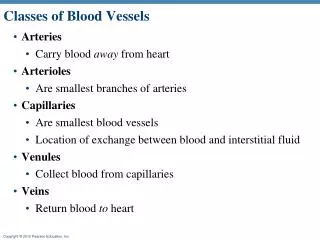
Classes of Blood Vessels. Arteries Carry blood away from heart Arterioles Are smallest branches of arteries Capillaries Are smallest blood vessels Location of exchange between blood and interstitial fluid Venules Collect blood from capillaries Veins Return blood to heart.

E N D
Classes of Blood Vessels • Arteries • Carry blood away from heart • Arterioles • Are smallest branches of arteries • Capillaries • Are smallest blood vessels • Location of exchange between blood and interstitial fluid • Venules • Collect blood from capillaries • Veins • Return blood to heart
Vascular Components Oxygenated blood from heart Deoxygenated blood to heart Valve Connective tissue Smooth muscle Endothelium Lumen Microcirculation Arteriole Venule Network of capillaries Artery Vein Basement membrane Endothelium Capillary
Generalized Structure of Blood Vessels • Arteries and veins are composed of three tunics • tunica interna, • tunica media • tunica externa • Capillaries are composed of endothelium with sparse basal lamina
Structure of vessel walls • The walls of blood vessels are too thick to allow diffusion between blood stream and surrounding tissues or the tissues of the blood vessels. • The walls of large vessels contain small blood vessels that supply both tunica media and externa – vasa vasorum
Capillaries • Capillaries are the smallest blood vessels • Walls consisting of a thin tunica interna, one cell thick • Allow only a single RBC to pass at a time • Pericytes on the outer surface stabilize their walls • There are three structural types of capillaries: continuous, fenestrated, and sinusoids
Continuous Capillaries • Continuous capillaries are abundant in the skin and muscles • Endothelial cells provide an uninterrupted lining • Adjacent cells are connected with tight junctions • Intercellular clefts allow the passage of fluids • Continuous capillaries of the brain: • Have tight junctions completely around the endothelium • Constitute the blood-brain barrier
Fenestrated Capillaries • Found wherever active capillary absorption or filtrate formation occurs (e.g., small intestines, endocrine glands, and kidneys) • Characterized by: • An endothelium riddled with pores (fenestrations) • Greater permeability than other capillaries
Sinusoids • Highly modified, leaky, fenestrated capillaries with large lumens • Found in the liver, bone marrow, lymphoid tissue, and in some endocrine organs • Allow large molecules (proteins and blood cells) to pass between the blood and surrounding tissues
Capillary Beds • Vascular shunts – Metarteriole--is a vessel that emerges from an arteriole, passes through the capillary network and empties into a venule. • Proximal portions are surrounded by scattered smooth muscle cells whose contraction and relaxation help regulate the amount and force of the blood. • Distal portion has no smooth muscle fibers and is called a thoroughfare channel. • True capillaries – 10 to 100 per capillary bed, capillaries branch off the metarteriole and return to the thoroughfare channel at the distal end of the bed • At their site of origin, there is a ring of smooth muscle fibers called a precapillary sphincter that controls the flow of blood entering a true capillary
Capillary Beds Figure 19.4a
Capillary Beds Figure 19.4b
Blood Flow • The purpose of cardiovascular regulation is to maintain adequate blood flow through the capillaries to the tissues • Actual volume of blood flowing through a vessel, an organ, or the entire circulation in a given period: • Is measured in ml/min. • Is equivalent to cardiac output (CO), considering the entire vascular system • Is relatively constant when at rest • Varies widely through individual organs
Blood Flow Through Tissues • Blood flow, or tissue perfusion, is involved in: • Delivery of oxygen and nutrients to, and removal of wastes from, tissue cells • Gas exchange in the lungs • Absorption of nutrients from the digestive tract • Urine formation by the kidneys • Blood flow is precisely the right amount to provide proper tissue function
Circulatory Shock • Circulatory shock – any condition in which blood vessels are inadequately filled and blood cannot circulate normally • Results in inadequate blood flow to meet tissue needs • Three types include: • Hypovolemic shock – results from large-scale blood loss or dehydration • Vascular shock – normal blood volume but too much accumulate in the limbs (long period of standing/sitting) • Cardiogenic shock – the heart cannot sustain adequate circulation
Blood flow • Capillary blood flow is determined by the interplay between: • Pressure • Resistance • The heart must generate sufficient pressure to overcome resistance
Physiology of Circulation: Blood Pressure (BP) • Force per unit area exerted on the wall of a blood vessel by its contained blood • Expressed in millimeters of mercury (mm Hg) • Measured in reference to systemic arterial BP in large arteries near the heart • The differences in BP within the vascular system provide the driving force that keeps blood moving from higher to lower pressure areas • Absolute pressure is less important than pressure gradient
Physiology of Circulation: Resistance • Opposition to flow • Measure of the amount of friction blood encounters • Generally encountered in the peripheral systemic circulation • Because the resistance of the venous system is very low (why?) usually the focus is on the resistance of the arterial system: • Peripheral resistance (PR) is the resistance of the arterial system
Physiology of Circulation: Resistance • Three important sources of resistance • Blood viscosity • Total blood vessel length • Blood vessel diameter
Resistance – constant factors • Blood viscosity • The “stickiness” of the blood due to formed elements and plasma proteins • Blood vessel length • The longer the vessel, the greater the resistance encountered
Resistance – frequently changed factors • Changes in vessel diameter are frequent and significantly alter peripheral resistance • Small-diameter arterioles are the major determinants of peripheral resistance • Frequent changes alter peripheral resistance • Fatty plaques from atherosclerosis: • Cause turbulent blood flow • Dramatically increase resistance due to turbulence
Systemic Blood Pressure • The pumping action of the heart generates blood flow through the vessels along a pressure gradient, always moving from higher- to lower-pressure areas • Pressure results when flow is opposed by resistance • Systemic pressure: • Is highest in the aorta • Declines throughout the length of the pathway • Is ~0 mm Hg in the right atrium • The steepest change in blood pressure occurs between arteries to arterioles
Arterial Blood Pressure • Arterial pressure is important because it maintains blood flow through capillaries • Blood pressure near the heart is pulsatile • Systolic pressure: pressure exerted during ventricular contraction • Diastolic pressure: lowest level of arterial pressure
Arterial Blood Pressure • A pulse is rhythmic pressure oscillation that accompanies every heartbeat • Pulse pressure = difference between systolic and diastolic pressure • Mean arterial pressure (MAP): pressure that propels the blood to the tissues MAP = diastolic pressure + 1/3 pulse pressure • Pulse pressure and MAP both decline with increasing distance from the heart • Efficiency of the circulation can be assessed by taking pulse and blood pressure measurements
Factors that Influence Mean Arterial Pressure Figure 15-10
How increases in cardiac output and total peripheral resistance increase mean arterial pressure
Alterations in Blood Pressure • Hypotension – low BP in which systolic pressure is below 100 mm Hg • Hypertension – condition of sustained elevated arterial pressure of 140/90 or higher • Transient elevations are normal and can be caused by fever, physical exertion, and emotional upset • Chronic elevation is a major cause of heart failure, vascular disease, renal failure, and stroke
Capillary Blood Pressure • Ranges from 15 to 35 mm Hg • Low capillary pressure is desirable • High BP would rupture fragile, thin-walled capillaries • Low BP is sufficient to force filtrate out into interstitial space and distribute nutrients, gases, and hormones between blood and tissues • Capillary blood pressure is one of the driving forces for transport through the capillary wall (filtration)
Exchange of materials across the wall of a capillary Endothelial cell Pores Plasma Interstitial fluid O2, CO2, fatty acids, Steroid hormones Lumen Diffusion through cells (lipid-soluble substances) Diffusion through pores (water-soluble substances) Na+, K+, glucose Capillary Restricted movement of most plasma proteins (cannot cross capillary wall) Proteins Transcytosis of exchangeable proteins across cells Water-filled pore cell Endothelial Cytoplasm Plasma membrane
Net Filtration Pressure (NFP) • NFP—comprises all the forces acting on a capillary bed • NFP = (HPc—HPif)—(OPc—OPif) • At the arterial end of a bed, hydrostatic forces dominate • At the venous end, osmotic forces dominate • Excess fluid is returned to the blood via the lymphatic system
Hydrostatic Pressures • Capillary hydrostatic pressure (HPc) (capillary blood pressure) • Tends to force fluids through the capillary walls • Is greater at the arterial end (35 mm Hg) of a bed than at the venule end (17 mm Hg) • Interstitial fluid hydrostatic pressure (HPif) • Usually assumed to be zero because of lymphatic vessels
Colloid Osmotic Pressures • Capillary colloid osmotic pressure (oncotic pressure) (OPc) • Created by non-diffusible plasma proteins, which draw water toward themselves • ~26 mm Hg • Interstitial fluid osmotic pressure (OPif) • Low (~1 mm Hg) due to low protein content
HP =hydrostatic pressure • Due to fluid pressing against a wall • “Pushes” • In capillary (HPc) • Pushes fluid out of capillary • 35 mm Hg at arterial end and 17 mmHg at venous end of capillaryin this example •In interstitial fluid (HPif) • Pushes fluid into capillary • 0 mm Hg in this example Arteriole Venule Interstitial fluid Capillary Net HP—Net OP (17—0)—(26—1) Net HP—Net OP (35—0)—(26—1) OP =osmotic pressure • Due to presence of nondiffusible solutes (e.g., plasma proteins) • “Sucks” •In capillary (OPc) • Pulls fluid into capillary • 26 mm Hg in this example •In interstitial fluid (OPif) • Pulls fluid out of capillary • 1 mm Hg in this example Net HP 35 mm Net OP 25 mm Net OP 25 mm Net HP 17 mm NFP (net filtration pressure) is 10 mm Hg; fluid moves out NFP is ~8 mm Hg; fluid moves in CHP – 35-17 mmHg IHP – 0 mmHg BCOP – 25 mmHg ICOP – 0 mmHg Figure 19.17
Fluid Exchange at a Capillary • Hydrostatic pressure and osmotic pressure regulate bulk flow Figure 15-19a
Venous Blood Pressure • Changes little during the cardiac cycle • Small pressure gradient, about 15 mm Hg
Factors Aiding Venous Return • Venous BP alone is too low to promote adequate blood return and is aided by the: • Respiratory “pump” – pressure changes created during breathing suck blood toward the heart by squeezing local veins • Muscular “pump” – contraction of skeletal muscles “milk” blood toward the heart • Valves prevent backflow during venous return
Controls of Blood Pressure • Short-term controls: • Counteract moment-to-moment fluctuations in blood pressure by altering peripheral resistance • Long-term controls regulate blood volume
Central Regulation Central regulation involves neuroendocrine mechanisms that control the total systemic circulation. This regulation involves both the cardiovascular centers and the vasomotor centers. Short-term elevation of blood pressure bysympatheticstimulation of theheart and peripheralvasoconstriction Stimulation ofreceptors sensitiveto changes insystemic bloodpressure orchemistry Activation ofcardiovascularcenters Neuralmechanisms Stimulationof endocrineresponse Long-term increasein blood volumeand blood pressure Endocrine mechanisms If autoregulation is ineffective HOMEOSTASISRESTORED
Short-Term Mechanisms to controls of Blood Pressure • Autoregulation • Causes immediate, localized homeostatic adjustments • Neural mechanisms • Respond quickly to changes at specific sites
Autoregulation of blood flow within tissues • Autoregulation – automatic adjustment of blood flow to each tissue in proportion to its requirements at any given point in time • Local vasodilators accelerate blood flow in response to: • Decreased tissue O2 levels or increased CO2 levels • Generation of lactic acid • Rising H+ concentrations in interstitial fluid • Local inflammation • Elevated temperature • Vasoconstrictors: • Injured vessels constrict strongly (why?) • Drop in tissue temperature (why?)
Myogenic (myo =muscle; gen=origin) Controls • Inadequate blood perfusion (tissue might die) or excessively high arterial pressure (rupture of vessels) may interfere with the function of the tissue • Vascular smooth muscle can prevent these problems by responding directly to passive stretch (increased intravascular pressure) • The muscle response is by resist to the stretch and that results in vasoconstriction • The opposite happens when there is a reduced stretch • The myogenic mechanisms keeps the tissue perforation relatively constant.
Short-Term Mechanisms: Neural Controls • Neural controls of peripheral resistance: • Alter blood distribution in response to demands • Maintain MAP by altering blood vessel diameter • Neural controls operate via reflex arcs involving: • Vasomotor centers and vasomotor fibers • Baroreceptors • Vascular smooth muscle
Short-Term Mechanisms: Vasomotor Center • Vasomotor center – a cluster of sympathetic neurons in the medulla that oversees changes in blood vessel diameter • Maintains blood vessel tone by innervating smooth muscles of blood vessels, especially arterioles • Vasomotor activity is modified by: • Baroreceptors (pressure-sensitive) • chemoreceptors (O2, CO2, and H+ sensitive) • bloodborne chemicals
Short-Term Mechanisms: Baroreceptor-Initiated Reflexes Carotid bifurcation Carotid sinus Common carotid artery Arterial baroreceptors Aortic arch
Short-Term Mechanisms: Baroreceptor-Initiated Reflexes • Increased blood pressure stimulates the cardioinhibitory center to: • Increase vessel diameter • Decrease heart rate, cardiac output, peripheral resistance, and blood pressure • Declining blood pressure stimulates the cardioacceleratory center to: • Increase cardiac output and peripheral resistance • Low blood pressure also stimulates the vasomotor center to constrict blood vessels