Download
1 / 26
260 likes | 679 Vues
ANATOMY OF THE FEMALE PELVIC FLOOR AND PERINEUM Dr. A. O. FAWOLE DEPT. OF OB-GYN UCH. Pelvic Floor and Perineum. Anterior perineum Posterior perineum Pelvic Floor. Perineum. Borders of pelvic outlet Anteriorly – ischiopubic rami Posteriorly – coccyx and sacro-tuberous ligament

E N D
ANATOMY OF THE FEMALE PELVIC FLOOR AND PERINEUM Dr. A. O. FAWOLE DEPT. OF OB-GYN UCH
Pelvic Floor and Perineum • Anterior perineum • Posterior perineum • Pelvic Floor
Perineum • Borders of pelvic outlet • Anteriorly – ischiopubic rami • Posteriorly – coccyx and sacro-tuberous ligament • Division (ischial tuberosities) • Anterior perineum (urogenital triangle) • Posterior perineum (anal triangle) • Structures overly perineal membrane
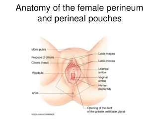
Structures of the Vulva • Mons • Labia • Clitoris • Vestibule • Erectile structures
Vulva • Mons • Hair-bearing skin • Cushion of adipose tissue • Labia majora • Hair-bearing skin • Termination of round ligaments of uterus • Labia minora • Hairless skin fold • Lacks bed of adipose tissue • Forms prepuce/frenulum of clitoris
Glands of the vulva • Labia majora – sebaceous gland • Sweat glands • Bartholin’s glands
Superficial compartment Space between subcutaneous tissues and perineal membrane • Clitoris • Crura • Vestibular bulb/Bartholin’s gland • Ischicavernosus (from ischial tuberosity) • Bulbospongiosus (from perineal body) • Superficial transverse perineal muscle • Muscles contained in superficial perineal pouch
The Perineal Membrane • Triagular sheet in anterior half of pelvic outlet • Dense fibromuscular tissue • Previously called urogenital diaphragm • Functions • Supports pelvic floor • Counteracts effects of raised intra-abdominal pressure • Counteracts effects of gravity
The Perineal Membrane • Complete sheet in males • Arises from ischiopubic rami • Attachments • Urethra • Vagina • Perineal body • Limits downward descent
Perineal Body • Mass of connective tissue • Synonym: central tendon of the perineum • Located between vagina and anus • Attachments • Perineal membrane • Superficial transverse perineal muscle • Bulbospongiosus • Levator ani • Coccyx (via external anal sphincter)
Posterior triangle • Anus • Anal sphincter • Levator ani • Ischio-rectal fossa
Ischio-rectal fossa • Lateral - Pudendal vessels/nerve (Pudendal canal) • Medial – Fascial covering of levator ani/external anal sphincter • Posterior – sacrotuberous ligament • Anterior – urogenital perineum • Floor – skin/ subcutaneous fat
Pudendal Nerve/Vessels • Similar channels • Nerve arises from sacral plexus (S2- S4) • Sensory • Motor • Artery: anterior division of internal iliac artery • Branches • Clitoral • Perineal (largest branch: muscles/skin) • Inferior rectal
Anal Sphincter • External anal sphincter • Superficial portion • Deep portion • Internal anal sphincter • Thickening of circular smooth muscle of anal wall • Important during repair of third (fourth) degree perineal laceration
The Pelvic Floor • Demands of upright posture • Support for pelvic structures • Passage of fetus • Openings for waste elimination • Fibromuscular floor • Visceral ligaments/fascia
Levator ani/Pelvic Wall • Levator ani most important muscle • Pubo-rectal • Pubo-coccygeal • Coccygeal • Origin: Posterior aspect of pubic bone, fascia of pelvic side wall, ischial spine • Insertion: Perineal body, anal sphincter, coccyx • Constitute pelvic diaphragm • Surfaces covered by fascia
Pelvic Floor • Pubo-rectal and pubo-coccygeal most medial • Pelvic diaphragm forms U-shaped layer of muscle • Opening end of U directed anteriorly (urogenital sinus) • Urethra • Vagina • rectum
Pelvic supports • Condensations of connective tissue or endopelvic fascia • Cardinal ligament • Uterosacral ligament • Pubocervical fascia
Clinical significance Prolapse of pelvic structures • Damage • Weakness
Cardinal ligament Transverse cervical/Mackenrodt’s ligament • Attaches lateral cervix/upper vagina to pelvic side wall • Connective tissue/involuntary muscle • Ureter in upper part
Uterosacral ligament • Postero-lateral attachment of lateral vaginal fornix/cervix/isthmus to sacro-iliac joint/third piece of sacrum
Pubo-cervical fascia • Extends between cardinal ligament and pubis • Lateral to the bladder • Significant in repair operation for cystocoele (anterior colporrhaphy)