Download
1 / 43
440 likes | 472 Vues
Explore the intricate layers of the skin and its appendages, including the mammary gland, to understand their functions, development, and composition. Learn about the epidermis, dermis, hypodermis, and the different skin appendages.

E N D
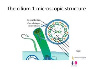
Lecture 10 General Medicine_3rd semester Microscopic structure of the skin Microscopic structure of skinappendages and mammary gland Development of the skin and skinappendages(derivatives)
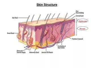
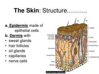
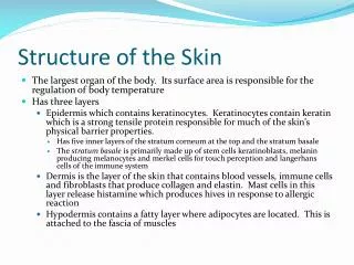
the skin (cutis) covers theexternal surface and is heaviest single organ of the human body -cca 16 % of total body weight - 1.2- 2.3 m2 in adults functions: • protects body against influence of the external factors, • contains tactile, cold, heat and pain receptors, • participates in immune response and processes, • is involved in regulation of body temperature, • serves as a complementary excretory organ (sebaceous and sudoriferous glands), • takes part in gaseous exchange (to a limited extent) the skin consists of • epidermis /an epithelial layer of ectodermic origin/ • dermis /a layer of connective tissue of mesenchymal origin/ and • hypodermis or subcutaneous tissue /a layer of loose connective tissue that may contain pads of adipose cells, the panniculus adiposus/ • skin appendages- either keratinised (hair and nails) or glandular (sebaceous and sudoriferous glands) the mammary gland is regarded as specialized and modified sweat gland (s)
EPIDERMIS is stratified squamouskeratinized epithelium cells are called keratinocytes it contains 3 less abundant cell types: melanocytes, Langerhans cells, and Merkel´s cells 2 types of the skin are distinguished: the thick (glabrous) skin - found on palms and soles the thin (hairy) skin - found elsewhere on the body surface /„thick“ and „thin“ refer to the thickness of the epidermis - it varies between 75 and 150 µm for the thin skin and 600-800 µm for the thick skin/
5.Stratum corneum:lies at the surface, consists of 15-20 layers of flattened non- nucleated keratinised cells whose cytoplasm is filled witha birefringent filamentous scleroprotein – keratin 4. Stratum lucidum : translucent and thin it lacks regularly in the thin skin; the layer contains dead, anucleated and eosinophilic cells,desmosomes are still evident 3. Stratum granulosum: 3 -5 layers of flattened polygonal cells with keratohyalin granulesand membrane-coated lamellar granules(composed of lamellar discs formed by lipid bilayers) 2.Stratum spinosum: consists of cuboidal, polyhedral, or slightly flattened cells, the cytoplasm projects into processes that are filled with bundles of tonofilaments(under light microscope as tonofibrils) cells in mitoses both strata are called by common term as stratum germinativum 1. Stratum basale(stratum cylindricum): a single layer of basophilic columnar or cuboidal cells, contain cytokeratin filaments numerous desmosomes, intense mitotic activity
3. Stratum granulosum 2.Stratum spinosum: 1. Stratum basale
After keratinisation, the cells consist of only fibrillar and amorphous proteins and thickened plasma membranes called horny cells Melanocytes are specialized cells of the epidermis located beneath or between cells of the stratum basale and in hair follicles cells synthesize and produce eumelanin synthesized melanin granules are then injected in the cytoplasm of keratinocytes
the color of the skin results from the content of melanin supranuclear location of granules
Langerhans cells (dendritic cells) are star shaped cells, found mainly in the stratum spinosum of the epidermis 2 - 8 % of the epidermal cells cells are supposed for the bone-marrow-derived macrophagesand are capable of binding and presenting antigens toT lymphocytes Merkel´s cells occur mostly in the thick skin they resemble keratinocytes but contain small dense granules in the cytoplasm numerous nerve endings terminate at the base of each Merkel´s cell - sensory mechanoreceptors
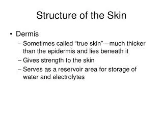
DERMIS it supports the epidermis and reaches the thickness about 4 mm 2 layers with rather indistinct boundaries: • the outermost papillary layer (stratum papillare corii ) - of loose connective tissue with networks of elastic and reticular fibers • the deeper reticular layer (stratum reticulare corii) - of irregular dense connective tissue (collagen I) the principal glycosaminoglycan of the dermis is dermatan sulfate thepapillary layer the reticular layer
in the thick (glabrous) skin found onpalms and solesdermal papillae run in pairs they are suppported with common corial ridges corial ridges correspond to thecristae cutis limited by sulci cutis on the surface of the epidermis orientation of cristae cutis is unique for each individual dactyloscopy
the dermis has a rich network of blood andlymph vessels blood may pass through capillaries or directly from arteries toveins via arteriovenous anastomoses or shunts these play a very important role in temperature and bloodpressure regulation
nerve supply of the skin is very rich SUBCUTANEOUS TISSUE (hypodermis, telasubcutanea) consists of loose connective tissue that binds the skin loosely to the subjacent organs (muscles, perichondrium or periosteum) it may contain fat cells
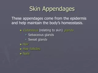
Skin derivatives of glandular type glands of the skin are of 2 types: • sweat glands • sebaceous glands Sweat glands- widely distributed throughout the body, are specialized for production of sweat that cools the body by evaporation, and other complex secretions eccrine sweat glands(gll. sudoriparae, gll. globiformes) are found everywhere on the body surface except the free margin of the lip, the prepuce and glans penis, the clitoris, and labia minora glandsare simple, coiled tubular consist of: the secretory portion the duct
the secretory portion: the thick basement membrane, myoepithelial cells and secretory cells: dark cells (mucoid cells) are pyramidal cells with basophilic cytoplasmgranules contain glycoproteins clear cells are devoid secretory granules but contain an abundance of glycogen particles the duct: a)properduct - in the dermis, its wall is composed of two-layered epithelium, myoepithelial cells and the basement membrane b)intraepidermal canaliculus - located in the epidermis it has no proper wall
secretion of eccrine glands is not viscous and contains little protein, components are water, sodium chloride, urea, ammonia, and uric acid
apocrine sweat glands in the axillaryand anal regions the secretory partthat is much larger than that in eccrine sweat glands and is lined by a cuboidal or columnar eosinophilic secretory cells theductthat opens into hair follicle glands produce a viscous milky white odorous secretion apocrine glands startsecretion after puberty, underinfluence of sex steroids modified apocrine sweat glands: the ceruminous glands in the external auditory meatus, glands of Montgomery in the nipple, and glands of Moll in eyelids
Sebaceous glands are holocrine type and are associated with hair follicles they occur practically on all body surfaces except the palms of the hands and soles of the feet consists of several alveoli (acini) and short duct that opens in the upper portion of a hair follicle and is lined by a stratified squamous epithelium
acini are composed of two types of cells: an outer layer of stem cells called basal cells and central group of cells that accumulate lipid droplets in their cytoplasm central cells disintegrate and become a part of sebum (holocrine secretion) sebaceous glands begin to function at puberty
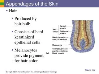
Skin derivatives ofkeratinizedtype hairs, nails hairs - elongated keratinised fibers derived from invaginations of epidermal epithelium, occur everywhere on the body except the palms, soles, lips, glans penis, clitoris, andlabia minora hair consists of - free part(scapus pili) and - hair root (radix pili) - an extendedpart of the hair root is hair bulb - hair papilla formed by vascularized loose connective tissue -hair follicle
Components of the hair • the hair medulla - central part consisting of 2 to 3 rows (columns) of lightly stained, polyhedral cells, it is discernible only in the hair bulb and in thick hairs, • the hair cortex - several layers of spindle-shaped (fusiform), tightly packed cells with melanin granules, • the hair cuticle (epidermicule) - is a layer of single keratinised cells that overlap each other and whose the ends are directed towards the scapus pili Hair follicle envelopes the hair root • the internal root sheath -lies close the hair and comprises three layers: Henle's and Huxley's layers, which contain eosinophilic trichohyaline granules, and the cuticle of keratinised cells; the internal root sheath grows from the hair bulb and its cells gradually keratinise towards the neck of the hair follicle (the neck = region of the opening of sebaceous gland) • the external root sheath - is continuous with the germinal epidermal layer and thins towards the hair bulb; it consists of lightly stained cells and the basement membrane • the connective tissue sheath - thin leaf of dense collagen tissue that links the hair to its surrounding tissue musculus arrector pili or arrector pili muscle is a bundle of smooth muscle cells stretched between the lower portion of the hair follicle and the superficial layer of dermis at the side where the loose hair end makes a sharp angle with the epidermis contraction of the arrector pili muscle causes a depression of the skin where the muscles attach to the dermis. This produces the "gooseflesh"
NAILS plates of keratinised epithelial cells on dorsal aspect of distal phalanxes (phalanges) • the proximal part of the nail, hidden in the nail groove, is thenail root • the nail root is covered with eponychium (or cuticle) • the nail plate- corresponds to the stratum corneum of the skin • rests on a bed of epidermis termed the nail bed- is composed of stratum basale and the stratum spinosum The cells of the nail bed that are under the root of the nail constitute the matrix.
MAMMARY GLANDS are modified apocrine sweat glands producing milk each mammary gland consists of 15-25 lobes which are separated each others by dense connective tissue lobes are drained with excretory lactiferous ducts that open in the nipple (15-25 openings) histological structure of mammary glands varies according to sex, age, and physiologic status Nonlactating mammary glands glandular tissue is reduced on only duct system, e.g. lactiferous ducts, terminal interlobular ducts and intralobular ducts an area of one interlobular duct is a lobule the lobules are separated by a denser, less cellular interlobular connective tissue spaces within lobules are filled with loose intralobular tissue abundant in cells.
Gl. mammae mamma non lactans
Lactating mammary glands glandular tissue is fully differentiated by thin connective tissue septa, the glandular parenchyma is divided into the lobules lobules contains spherical to elongated acini (alveoli) differ in size the wall of acini consist of - the basement membrane - cuboidal or columnar secretory cellsand - myoepithelial cells located between the basement membrane and bases of secretory cells ducts a) intralobular ducts lined by a simple cuboidal to columnar epithelium b) lactiferous ducts lined by two layers of columnar cells which, in the lactiferous sinus, changes into stratified squamous epithelium • the first secretion appearing after birth is called the colostrum • it contains less fat and more protein than regular milk and is rich in antibodies
Gl. mammae mamma lactans
DEVELOPMENT OF SKIN AND SKIN APPENDAGES
Epidermis initially, a single layer of ectodermal cells covers the embryo starting from the 2nd month, the ectodermal cells divide and form a superficial protective layer offlattened cells, the periderm or epitrichium at the end of 4th month, the epidermis acquires its definitive arrangement and 4 layers are distinguished: basal, spinous, granular and horny layer
cells that have been exfoliated during fetal life form part of the vernix caseosa, a white, cheese-like, protective substance that covers the fetal skin during the early fetal period, melanoblasts migrate from the neural crest to the dermoepidermal junction, where they differentiate into melanocytes
Dermis the dermis is derived from the mesenchyme underlying the surface ectoderm the mesenchyme arises from 2 sources: from the somatic layer of lateral mesoderm (most of the mesenchyme), from the dermomyotome regions of the somites (in lesser extent) by 11 weeks, the mesenchymal cells have begun to produce collagenous and elastic connective tissue fibers as the epidermal ridges form, the dermis project upward into the epidermis and forms dermal papillae
Eccrine sweat glands develop as solid epidermal downgrowths that extendinto the underlying dermis as buds elongate, their ends become coiled, forming the primordia of future secretory portions of glands
Development of hairs they develop early in the fetal period, but they do not become readily visibleuntil about 20th week first recognizable hairs occur on the eyebrows, upper lip, and chin a hair follicle begins to develop as a solid downgrowth of the stratum germinativun of the epidermis called the hair bud it extends intothe underlying dermis the deepest part of the hair bud soon becomes club-shaped, forming ahair bulb the epithelial cells of the centre of the hair bulb constitute the germinal matrix - it gives rise to properhair the hair bulb is then invaginated by a small mesenchymal hair papilla
the peripheral cells of the developing hair bud (follicle) form the epithelial root sheath the surrounding mesenchymal cells differentiate into the dermal (connective tissue) root sheath the first hairs are called lanugo, are fine and colourless lanugo is replaced during the perinatal period by coarser hairs, called vellus these hairs persist over most of the body, except in the axillary and pubic regions hairs of those regions are replaced during puberty
apocrine sweat glands (axilla, pubic region, anal region, areolae) develop from the hair follicle as sebaceous glands
Development of mammary gland mammary glands develop during the 6th week as a solid downgrowth of theepidermis that grow against the underlying mesenchyma downgrowths occur along the mammary ridges - two thickened strips of ectoderm that run from the axillary to the inguinal regions in human embryos, mammary ridges occur during the 4th week, but except the pectoral area rapidly disappear each primary mammary budsoon gives rise to severalsecondary buds that develop into lactiferous ducts and their branches the fibrous connective tissue and fat develop from the surrounding mesenchyme
Nails toenails and fingernails begin to develop at the distal ends of digits at about 10 weeks, development of fingernailsprecedes that of the toenails the nails first appear as thickened areas of the developing epidermis on the dorsal aspect of each digit the nail fieldsare surrounded laterally and proximally by folds - nail folds cells from the proximal nail fold grow over the nail field and become keratinised to form the nail, or nail plate at first, superficial layers of epidermis called the eponychium cover the developing nail, this later degenerates, except at thebase of the nail, where it persists