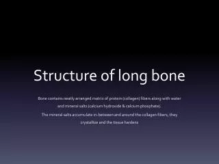
Microscopic Structure of Bone
Microscopic Structure of Bone. Osseous Tissue. Another name for bone tissue Bone is a connective Tissue Widely spread cells Matrix: Water, Collagen Fibers, Mineral Salts. Calcification. Hardening of bone tissue by the deposition of mineral salts in the collagen fiber of the matrix.

Microscopic Structure of Bone
E N D
Presentation Transcript
Osseous Tissue • Another name for bone tissue • Bone is a connective Tissue • Widely spread cells • Matrix: • Water, Collagen Fibers, Mineral Salts
Calcification • Hardening of bone tissue by the deposition of mineral salts in the collagen fiber of the matrix
Hardness and Flexibility • Hardness – Provided by the crystallized mineral salts • Flexibility – Provided by the collagen fibers • Bones can resist being stretched or torn apart
Cells • There are 4 major types of cells found in osseous tissue • Osteoblast • Osteocyte • Osteoprogenitor • Osteoclast
Osteoblast • Bone building cells • Synthesize and secrete collagen fibers and other organic components needed to build the matrix of the tissue • Osteoblasts surround themselves with matrix, become trapped in their secretions and become osteocytes • Do not undergo Mitosis
Osteocyte • Mature Bone Cell • Main cells in bone tissue • Maintains daily metabolism • Do not undergo mitosis
Osteoprogenitor • “Bone stem cells” • These cells undergo mitosis then differentiate to form osteoblasts
Osteoclasts • HUGE cells formed from the fusion of as many as 50 monocytes • Concentrated in the endosteum • Release lysosomal enzymes and acids to digest the matrix
Reabsorption • Breakdown of bone matrix • Part of the normal development, growth, repair and maintenance
Categories of Bone Tissue • Bone has many small spaces between the cells and matrix – it is not completely solid. • The category of tissue is based on the size and distribution of these spaces • About 80% of bone is compact bone; 20% is spongy bone
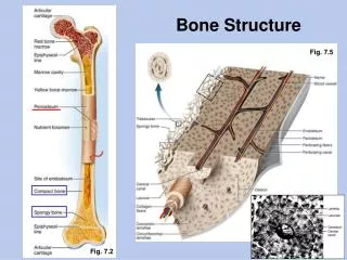
Compact Bone Tissue • Contains very few spaces • Forms the external layer of all bones and the diaphyses of long bones • Provides protection and support • Resists stress produced by weight and movement
Osteon • Organizational Unit of Compact Bone
Perforating Canals • Transverse openings through which vessels from the periosteum penetrate the compact bone and eventually meet up with other vessels
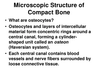
Central (Haversian) Canal • Run longitudinally through bone • It is the center of the osteon • Contains blood vessels and nerves
Concentric Lamellae • Rings of hard calcified matrix surrounding the central canal
Lacunae • Means “little lake” • Small spaces between the lamellae • Contain osteocytes
Canaliculi • Minute canals that radiate off the lacunae in all directions. • Contain projections of the osteocytes • Connect lacunae creating a network throughout the compact bone to provide nutrients and oxygen to all the osteocytes and to get rid of waste
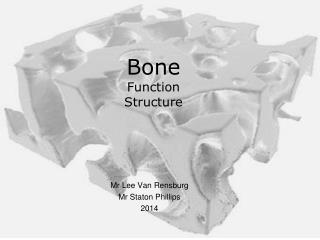
Does not contain ostons • Made of trabeculae – an irregular network of thin columns of bone with many spaces in between • Trabeculae contain osteocytes within lacunae connected by canaliculi
Spongy tissue makes up most of flat, short and irregular bones • Forms most of the epiphyses of long bones • Found in a narrow rim around the medullary cavity
Spongy tissue is light – reducing the weight of the skeletal system • Red bone marrow is found in the spaces between trabeculae • Hemopoiesis only occurs in the hip bones, ribs, sternum, vertebrae and epiphyses of long bones – where red bone marrow is found
Bone Scan • Radioactive Tracer is injected through an IV and absorbed by the bone • A scanning device measures the amount of radiation emitted by the bones and translates the information into an x-ray • Normal bones have a consistent gray color • Darker/lighter areas indicate an abnormality • Ex - Bone cancer, abnormal healing, infections, arthritis
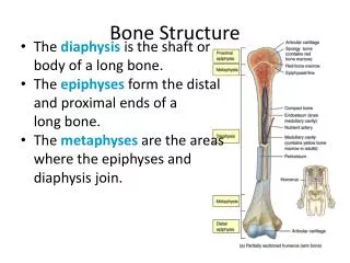
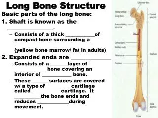
Checkpoint Questions – answer these in your notes… • What kinds of tissue make up the skeletal system? • How do red and yellow bone marrow differ in composition and function? • What are the types of bones? • Draw and label the parts of a typical long bone. • What are the 4 types of bone cells? • How are spongy and compact bone different in microscopic appearance, function and location?