Download
1 / 13
150 likes | 717 Vues
Learn about osteocytes, Haversian system, bone types, axial and appendicular skeleton divisions, and key bone terminology for comprehensive knowledge.

E N D
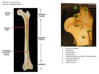
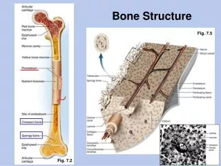
Microscopic Structure of Compact Bone What are osteocytes? Osteocytes and layers of intercellular material form concentric rings around a central canal, forming a cylinder-shaped unit called an osteon (Haversian system). Each central canal contains blood vessels and nerve fibers surrounded by loose connective tissue.
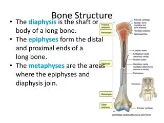
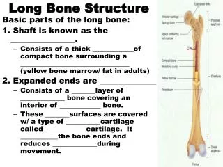
Label Femur Diagram Handout • Review the following terms: • Distal • Proximal
Bone types Remember: Bones differ greatly in size and shape, but are similar in structure. Long Bones – work as levers; EX: bones of upper and lower limbs Short Bones – short and cubed shaped; found in wrist and ankles Flat Bones - broad surfaces for protection of organs and attachment of muscles; EX: ribs, cranial bones Irregular Bones – vertebrae and some bones in skull
Skeletal Organization The skeleton is divided into two major parts: Axial (???) skeleton Appendicular (???) skeleton
Axial Skeleton Consists of the bony and cartilage portions that support and protect the organs of the head, neck, and trunk.
Axial Skeleton parts: Skull – made of the cranium and facial bones Hyoid bone – found in the neck between the lower jaw and the larynx (this bone supports the tongue) Vertebral column – vertebrae, sacrum, and coccyx Thoracic cage – ribs (12 pair) and sternum
Appendicular Skeleton Consists of bones of the upper and lower limbs and the bones that anchor the limbs to the axial skeleton.
Appendicular Parts: Pectoral Girdle – scapula and clavicle Upper Limbs – humerus, radius ulna, carpals, metacarpals, phalanges Pelvic Girdle – pelvis Lower Limbs – femur, tibia, fibula, patella, tarsals, metatarsals, phalanges
Which portion, axial or appendicular, do you think would have the largest number of bones? Table 7.1, p.137, shows the skeletal organization in a semi-outline format. ***Important bone terminology: Table 7.2, p.137
Skeletal Diagram Label together Use 4 different colored highlighters to highlight by TYPE of bone (long, short, flat, irregular)