Download
1 / 41
410 likes | 619 Vues
Appendages of the Skin. Derivatives of the epidermis Sweat glands Oil glands Hairs and hair follicles Nails. Sweat Glands. Two main types of sweat (sudoriferous) glands Eccrine (merocrine) sweat glands—abundant on palms, soles, and forehead

E N D
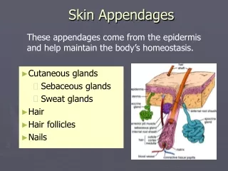
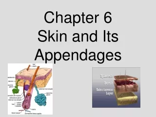
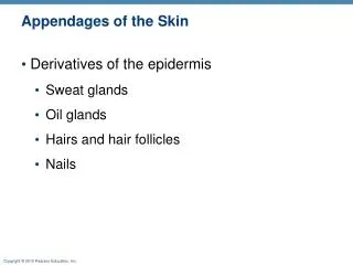
Appendages of the Skin • Derivatives of the epidermis • Sweat glands • Oil glands • Hairs and hair follicles • Nails
Sweat Glands • Two main types of sweat (sudoriferous) glands • Eccrine (merocrine) sweat glands—abundant on palms, soles, and forehead • Sweat: 99% water, NaCl, vitamin C, antibodies, dermcidin, metabolic wastes • Ducts connect to pores • Function in thermoregulation
Sweat pore Eccrine gland Sebaceous gland Duct Dermal connective tissue Secretory cells (b) Photomicrograph of a sectioned eccrine gland (220x) Figure 5.5b
Sweat Glands • Apocrine sweat glands—confined to axillary and anogenital areas • Sebum: sweat + fatty substances and proteins • Ducts connect to hair follicles • Functional from puberty onward (as sexual scent glands?) • Specialized apocrine glands • Ceruminous glands—in external ear canal; secrete cerumen • Mammary glands
Sebaceous (Oil) Glands • Widely distributed • Most develop from hair follicles • Become active at puberty • Sebum • Oily holocrine secretion • Bactericidal • Softens hair and skin
Sweat pore Sebaceous gland Dermal connective tissue Eccrine gland Sebaceous gland duct Hair in hair follicle Secretory cells (a) Photomicrograph of a sectioned sebaceous gland (220x) Figure 5.5a
Hair • Functions • Alerting the body to presence of insects on the skin • Guarding the scalp against physical trauma, heat loss, and sunlight • Distribution • Entire surface except palms, soles, lips, nipples, and portions of external genitalia
Hair • Consists of dead keratinized cells • Contains hard keratin; more durable than soft keratin of skin • Hair pigments: melanins (yellow, rust brown, black) • Gray/white hair: decreased melanin production, increased air bubbles in shaft
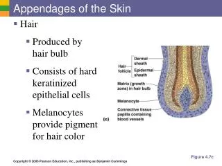
Follicle wall • Connective tissue root sheath • Glassy membrane • External epithelial root sheath • Internal epithelial root sheath Hair shaft Hair • Cuticle • Cortex • Medulla Arrector pili (a) Diagram of a cross section of a hair within its follicle Sebaceous gland Hair root Hair bulb Figure 5.6a
Follicle wall • Connective tissue root sheath • Glassy membrane • External epithelial root sheath • Internal epithelial root sheath Hair • Cuticle • Cortex Hair shaft • Medulla Arrector pili (b) Photomicrograph of a cross section of a hair and hair follicle (250x) Sebaceous gland Hair root Hair bulb Figure 5.6b
Hair Follicle • Extends from the epidermal surface into dermis • Two-layered wall: outer connective tissue root sheath, inner epithelial root sheath • Hair bulb: expanded deep end
Hair Follicle • Hair follicle receptor (root hair plexus) • Sensory nerve endings around each hair bulb • Stimulated by bending a hair • Arrector pili • Smooth muscle attached to follicle • Responsible for “goose bumps”
Hair shaft Arrector pili Sebaceous gland Follicle wall Hair root • Connective tissue root sheath Hair bulb • Glassy membrane • External epithelial root sheath • Internal epithelial root sheath Hair root • Cuticle • Cortex • Medulla Hair matrix Hair papilla Melanocyte Subcutaneous adipose tissue (c) Diagram of a longitudinal view of the expanded hair bulb of the follicle, which encloses the matrix Figure 5.6c
Follicle wall • Connective tissue root sheath Hair shaft • Glassy membrane • External epithelial root sheath • Internal epithelial root sheath Arrector pili Hair root • Cuticle Sebaceous gland • Cortex • Medulla Hair root Hair matrix Hair bulb Hair papilla Subcutaneous adipose tissue (d) Photomicrograph of longitudinal view of the hair bulb in the follicle (160x) Figure 5.6d
Types of Hair • Vellus—pale, fine body hair of children and adult females • Terminal—coarse, long hair of eyebrows, scalp, axillary, and pubic regions (and face and neck of males)
Types of Hair • Hair Growth • Growth phase (weeks to years) followed by regressive stage and resting phase (1–3 months) • Growth phase varies (6–10 years in scalp, 3–4 months in eyebrows)
Hair Thinning and Baldness • Alopecia—hair thinning in both sexes after age 40 • True (frank) baldness • Genetically determined and sex-influenced condition • Male pattern baldness is caused by follicular response to DHT
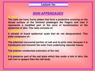
Structure of a Nail • Scalelike modification of the epidermis on the distal, dorsal surface of fingers and toes
Lateral nail fold Lunule (a) Free edge of nail Body of nail Eponychium (cuticle) Proximal nail fold Nail bed Root of nail Nail matrix (b) Hyponychium Phalanx (bone of fingertip) Figure 5.7
Functions of the Integumentary System • Protection—three types of barriers • Chemical • Low pH secretions (acid mantle) and defensins retard bacterial activity
Functions of the Integumentary System • Physical/mechanical barriers • Keratin and glycolipids block most water and water- soluble substances • Limited penetration of skin by lipid-soluble substances, plant oleoresins (e.g., poison ivy), organic solvents, salts of heavy metals, some drugs • Biological barriers • Dendritic cells, macrophages, and DNA
Functions of the Integumentary System • Body temperature regulation • ~500 ml/day of routine insensible perspiration (at normal body temperature) • At elevated temperature, dilation of dermal vessels and increased sweat gland activity (sensible perspirations) cool the body • Cutaneous sensations • Temperature, touch, and pain
Functions of the Integumentary System • Metabolic functions • Synthesis of vitamin D precursor and collagenase • Chemical conversion of carcinogens and some hormones • Blood reservoir—up to 5% of body’s blood volume • Excretion—nitrogenous wastes and salt in sweat
Skin Cancer • Most skin tumors are benign (do not metastasize) • Risk factors • Overexposure to UV radiation • Frequent irritation of the skin • Some skin lotions contain enzymes in liposomes that can fix damaged DNA
Skin Cancer • Three major types: • Basal cell carcinoma • Least malignant, most common • Squamous cell carcinoma • Second most common • Melanoma • Most dangerous
Basal Cell Carcinoma • Stratum basale cells proliferate and slowly invade dermis and hypodermis • Cured by surgical excision in 99% of cases
Squamous Cell Carcinoma • Involves keratinocytes of stratum spinosum • Most common on scalp, ears, lower lip, and hands • Good prognosis if treated by radiation therapy or removed surgically
Melanoma • Involves melanocytes • Highly metastatic and resistant to chemotherapy • Treated by wide surgical excision accompanied by immunotherapy
Melanoma • Characteristics (ABCD rule) A: Asymmetry; the two sides of the pigmented area do not match B: Border exhibits indentations C: Color is black, brown, tan, and sometimes red or blue D: Diameter is larger than 6 mm (size of a pencil eraser)
Burns • Heat, electricity, radiation, certain chemicals Burn (tissue damage, denatured protein, cell death) • Immediate threat: • Dehydration and electrolyte imbalance, leading to renal shutdown and circulatory shock
Partial-Thickness Burns • First degree • Epidermal damage only • Localized redness, edema (swelling), and pain • Second degree • Epidermal and upper dermal damage • Blisters appear
1st degree burn 2nd degree burn (a) Skin bearing partial thickness burn (1st and 2nd degree burns) Figure 5.10a
Full-Thickness Burns • Third degree • Entire thickness of skin damaged • Gray-white, cherry red, or black • No initial edema or pain (nerve endings destroyed) • Skin grafting usually necessary
3rd degree burn (b) Skin bearing full thickness burn (3rd degree burn) Figure 5.10b
Severity of Burns • Critical if: • >25% of the body has second-degree burns • >10% of the body has third-degree burns • Face, hands, or feet bear third-degree burns
Developmental Aspects: Fetal • Ectoderm epidermis • Mesoderm dermis and hypodermis • Lanugo coat: covering of delicate hairs in 5th and 6th month • Vernix caseosa: sebaceous gland secretion; protects skin of fetus
Developmental Aspects: Adolescent to Adult • Sebaceous gland activity increases • Effects of cumulative environmental assaults show after age 30 • Scaling and dermatitis become more common
Developmental Aspects: Old Age • Epidermal replacement slows, skin becomes thin, dry, and itchy • Subcutaneous fat and elasticity decrease, leading to cold intolerance and wrinkles • Increased risk of cancer due to decreased numbers of melanocytes and dendritic cells