Download
1 / 13
130 likes | 216 Vues
Lecture Ten. SKIN APPENDAGES A. Nails The nails are hard, horny plates that form a protective covering on the dorsal surface of the terminal phalanges the fingers and toes it represents a modified part of the zone of keratinization of the epidermis of skin. The nails consists of:

E N D
Lecture Ten SKIN APPENDAGES A. Nails The nails are hard, horny plates that form a protective covering on the dorsal surface of the terminal phalanges the fingers and toes it represents a modified part of the zone of keratinization of the epidermis of skin. The nails consists of: 1. Nail plate It consist of fused epidermal scale that do not desquamated. This plate composes of:- a. Nail body The attached uncovered portion of nail and its pink color because it is translucent and transmit the color from underlying vascular tissue. b. Free edge The anterior unattached extension of the nail. c. Nail root It is posterior part of the nail plate which lies under a fold of skin, the nail root is opaque than the nail body.
Lecture Ten 2. Nail bed Is the skin which lies under nail and reasting on nail. Consist of only the (stratum spinosum and basale) and the underlying dermis. 3. Nail wall It's a flod of skin which extends around the proximal and lateral borders of nail. 4. Nail groove Is the furrow between nail bed and nail wall. 5. Lunula Is the whitish part and the posterior end of nail. Represent the visible junction between body and root of nail plate. 6. Eponychium The stratum coneum extends distal upon the free surface of the nail. 7. Hyponychium The stratum corneum is thickened which is lying under the free border of the nail. 8. Germial matrix The germinative zone is thick near the nail root where it forms their germial matrix; the nail substance is formed by proliferation of cells in the germinal matrix.
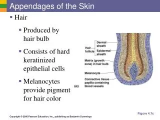
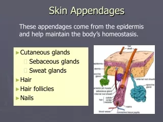
Lecture Ten B. Hairs Hairs are elongated keratinized structures derived from invagination of epidermal epithelium. Their color, size and disposition vary according to age, sex and region of the body. Hairs are found every where on the body except on the palms, soles, lips and glans penis and some few other sites. Each hairs has a free shaft and a root embedded in the skin, enclosing the hair root is a tubular invagination called hair follicle, which consist of epidermal and dermal portion, at is lower end the follicle expand into a hair bulb. Associated with the hair follicle are one or more sebaceous gland and bundle of smooth muscle fibers which formed the arrector pili, by its contraction it causes erection of the hair.
Lecture Ten Structure of the hair The hair consist of epidermal cells arranged in 3 concentric layers: 1. Medulla It forms the loose central axis and consist of (2-3) layers of shriken, cornified cuboidal cells, the cells often contain pigment. (Kertain). 2. Cortex Is composed of several layers of long flattened, pinle-shaped cells in which the keratin is of hard type. The pigment granules (melanin) found in the cells. The cells separated by air space, air accumulate in the intercellular spaces of both modularly and cortical responsible of modifies the hair color. 3. Hair cuticle The superficially layer composed of single layer of flat scale (cell with no nuclei) it is heavily keratinized.
Lecture Ten Structure of hair follicle It is consist of two sheaths: 1- Dermal root sheath, it is composed of 3 layers: a. The outer layer Is poorly defined and consist of bundle of collagen fibers and this correspond to the reticular layer of dermis. These fibers arrangement longitudinally. b. The middle layer Is thicker and consist of collagen fiber and correspond to papillary layer of dermis. These fibers arrangement circulatory. c. The inner or (glassy membrane) It is elongated the basal membrane consist of glycoprotein and corresponding to the basal lamina beneath epidermis.
Lecture Ten 2. Epidermal root sheath, this sheath divided two sub sheath. a. External root sheath It is thinner and composed of cells corresponding to the stratume germinativum of the epidermis. b. Internal root sheath It is a keratinized sheath enveloping the growing hair root and like the hair it is pushed up by addition of cells from the bulb. The internal sheath dose not extend above the point of entry of the duct of sebaceous gland into the follicle. It has 3 layers: * Henles layer It is directly in relation to the external root sheath composed of a single layer if flattened clear cells. * Huxley's layer It is consisting of several rows of elongated cells, the deeper cells contain nuclei but superficially nuclei are absent. * Cuticle of the internal root sheath It lies against the cuticle of the hair and is similar to it structure, which is consist of single layer of horny scales like cells.
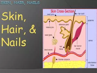
Lecture Ten The color of the hair Is due to melanin it is formed by melanocytes distributed in the matrix of the bulb of the follicle; there are only 3 colors of pigment. Black melanin derived form tyrosin. Brawn melanin derived form tyrosin. Pheno melanin derived form tryptophan. The variation in hair colors are combinations of different amount of the three pigments. Graying (whiting) of hair is due the loss of the pigment (melanocytes to form pigment). C. Glands of the skin 1. Sebaceous glands These glands are embedded in the dermis over most of the body surface; there are about 100 of these glands per square centimeter over most of the body, but the increases to (400-900/cm2) the face, forehead and scalp. Sebaceous glands which are not found in the palms and soles.
Lecture Ten The acinar glands that usually have several acini opening into a short duct, this duct usually ends in the upper portion of a hair follicle, the acini consist of a basal layer of undifferentiated flattened epithelial cells that rest on the basal lamina, these cells proliferate and differentiate filling the acini with rounded cells containing more fat droplets in their cytoplasm. Their nuclei gradually shrink and the cells become filled with fat droplets. The secretion of the sebaceous gland which is gradually moved to the surface of the skin. It is an example of a holocrine gland, because its product of secretion released with remnants of dead cells, this product a complex mixture of lipids that include triglycerides, waxes, squalene and cholesterol. Sebaceous glands begin to function at puberty, these glands development and growth are under the control of sex hormones. Before puberty, sebaceous glands are very small but at puberty in man their activity and size increase greatly under the influence of testosterone but in female the increased activity is caused by (Estrogen). When inflammation accure in these glands this result into Acne.
Lecture Ten 2. Sweat glands Sweat glands are tubular coiled glands distributed in the skin except upon the nail bed, margin of the lips and eardrum, they are most numerous in the palms and soles. The secretary portion is situated deeply in the dermis. The exceretory portion or duct rises to epidermis and spirals through it to reach the free surface where it opens by sweat pore.
Lecture Ten Structure of the sweat gland This gland composed of the: a. Secretary portion or unit This unit lined by a simple columnar or cuboidal epithelium supported by basal lamina and is surrounded by myoepithelial cell, contraction of these cells helps discharge the secretion. Two types of cells have been described in the secretory portion of merocrine sweat glands:- * Clear cells Are pyramidal in shape have a narrow apical area and a board base on basal lamina. The nucleus is round, the cytoplasm contains fat droplets and sometimes pigment granule also contain rough endoplasmic reticulum, and no secretary granules are present in cytoplasm by they have accumulation glycogen. * Dark cells (mucoid cells) Lined the lumen of the sweat gland secret mucus rich substance, it is shape resemble inverted cone, the broad end lined the lumen and the narrow end lined between adjacent clear cells and basal lamina. It has similar cytoplasmic organelles to those clear cells except it has glycoprotein containing secretary granules located in apical cytoplasm.
Lecture Ten b. Secretary duct Is continuous with secretary unit and narrows as it passes the epidermis to the surface. The wall of secretary duct made of (2 layers) of cuboidal epithelial cells of basal layer and cells of luminal layer, it doesn't have myoepithelial cells, the cells of inner layer of duct have microvilli when the duct reaches the epidermis it loses its wall and become a channel. It secret sweat contain (NaCl, H2O, uric acid, lactic acid and glucose).
Lecture Ten Type of sweat glands * Merocrine sweat glands It most common in all body except axilla and gentile area, their secretion occur by Exocytosis. * Apocrine sweat glands They are found in the axilla and areola of the nipple. They are larger sweat gland than merocrine sweat gland and produce a thicker secretion. Secretory cells are cuboidal to low columnar cells. Lumen of the gland is larger, the duct of the gland open into the upper portions of hair follicles (not on to skin surface) just superficial of entry of sebaceous glands. These large sweat gland show less coiling than merocrine sweat gland and myoepithelial cells are larger and form a more complete layer between the epithelial cells and basal lamina (no clear cells are present). The function of these glands are begin only at puberty. Functions of the sweat gland These glands play an important role in thermo regulation by providing moisture on the skin surface for evaporative cooling. They are responsive to nervous stress.