Download
1 / 64
651 likes | 1.04k Vues
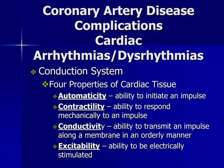
Coronary Artery Disease Complications Cardiac Arrhythmias/Dysrhythmias. Conduction System Four Properties of Cardiac Tissue Automaticity – ability to initiate an impulse Contractility – ability to respond mechanically to an impulse

E N D
Coronary Artery DiseaseComplicationsCardiac Arrhythmias/Dysrhythmias • Conduction System • Four Properties of Cardiac Tissue • Automaticity – ability to initiate an impulse • Contractility – ability to respond mechanically to an impulse • Conductivity – ability to transmit an impulse along a membrane in an orderly manner • Excitability – ability to be electrically stimulated
Cardiac Conduction System Specialized neuromuscular tissue • PR Interval: • SA Node – upper R atrium through Bachman’s Bundle • AV Node – internodal pathway • Bundle of His • QRS Complex: • Right and Left Bundle Branches • Purkinje Fibers
Calculating Heart Rate • EKG paper is a grid where time is measured along the horizontal axis. • Each small square is 1 mm in length and represents 0.04 seconds. • Each larger square is 5 mm in length and represents 0.2 seconds. • Voltage is measured along the vertical axis - 10 mm is equal to 1mV in voltage. • Heart rate can be easily calculated from the EKG strip: • Heart rate can be easily calculated from the EKG strip: • When the rhythm is regular: • the heart rate is 300 divided by the number of large squares between the QRS complexes. • e.g., if there are 4 large squares between regular QRS complexes, the heart rate is 75 (300/4=75). • The second method can be used with an irregular rhythm to estimate the rate: • Count the number of R waves in a 6 second strip and multiply by 10. • e.g., if there are 7 R waves in a 6 second strip, the heart rate is 70 (7x10=70).
Cardiac MonitoringCardiac Rhythm Analysis • Analyze the P waves – rate/rhythm • Analyze the QRS complexes – rate/rhythm • Determine the heart rate • Measure the PR Interval • Measure the QRS duration • Interpret the rhythm • Clinical significance? Hemodynamic status? • Appropriate Tx
Cardiac MonitoringNormal Sinus Rhythm • Atrial & Ventricular rhythms: regular • Rate: 60-100 beats/min • P waves: present consistent configuration, one P wave prior to each QRS complex • PR interval: .12 – .20 sec and constant • QRS duration: -.04 to .10 sec and constant
Cardiac MonitoringSinus Bradycardia • SA Node discharges < 60 beats/ min • Etiology: >parasympathetic stimulation / vagus nerve • Assess: LOC, Orientation, VS, PO, pain, escaped ventricular ectopy • Tx: If patient is symptomatic – raise legs up, move patient, Atropine – ACLS Bradycardia
Cardiac MonitoringSinus Tachycardia • SA Node discharge > 100 beats/ min • Etiology: Sympathetic stimulation – normal or abnormal response • Tx: Treat underlying cause • Cardiac Supply Problems • Cardiac Demand Problems • E.g., hypovolemia, hypoxemia, anxiety, pain, anemia, angina • Regular Narrow QRS - Adenosine
Sustained Tachy / Brady Dysrhythmias • Chest discomfort, or pain, radiation to jaw, back, shoulder or upper arm • Restlessness, anxiety, nervousness • Dizziness, syncope • Change in pulse strength, rate, rhythm • Pulse deficit • Shortness of breath, dyspnea • Tachypnea, Orthopnea • Pulmonary rales • S3 or S4 heart sounds • Jugular vein distention • Weakness, fatigue • Pale, cool skin, diaphoresis • Nausea, vomiting • Decreased urine output • Hypotension
Cardiac MonitoringParoxysmal Supraventricular Narrow QRS Tachycardia (PSVT) • SA Node rate 100-280 beats/min - Mean 170 beats/min • Etiology: Pre-excitation syndrome, e.g., Wolff-Parkinson White (WPW) Syndrome • Assess: Weakness, fatigue, chest pain, chest wall pain, hypotension, dyspnea, nervousness • Tx: Valsalva maneuvers: bearing down, gagging, ocular pressure, vomiting, carotid sinus massage, • Meds: Adenosine
Cardiac MonitoringAtrial Fibrillation • Most Common dysrhythmia in the US • Multiple rapid impulses from many atrial foci, rate of 350-600/min—depolarize the atrial in a disorganized and chaotic manner – atrial quiver • Results: • No P waves • No atrial contracts • No atrial kick • Irregular ventricular response
Cardiac MonitoringAtrial Fibrillation • Etiology: MI, RHD with Mitral Stenosis, CHF, COPD, Cardiomyopathy, Hyperthyroidism, Pulmonary emboli, WPW Syndrome, Congenital heart disease ** Mural Thrombi – increased risk for pulmonary & systemic thromboemboli to brain & periphery • Assess: VS, PO, Pulse Deficit, chest pain, syncope, hypotension • Symptoms worsen with increased ventricular response
Cardiac MonitoringAtrial Fibrillation • Tx: • TEE – Trans-esophageal echocardiogram • Identifies thrombi on valves • Medications to decrease the ventricular response - Metoprolol (Lopressor) • Oxygen • Prophylactic anticoagulation • Lovenox - Coumadin – long term • Cardioversion
Cardiac MonitoringAtrial Fibrillation • Tx: • Medications to decrease the ventricular response • Narrow QRS irreg rhythm–diltiazem; beta-blockers • Wide QRS reg rhythm – amiodarone • Wide QRS irreg rhythm – digoxin, diltiazem, verapermil, amiodarone • Oxygen • Prophylactic anticoagulation • Cardioversion
Cardiac MonitoringAtrial FibrillationCardioversion • Synchronized countershock • 50 – 100 Joules • Avoids delivering shock during repolarization • Patent intravenous line • Patient sedated – Versed • Oxygenation • ABC • Assess: VS, PO, Monitor cardiac rate - rhythm • Administer antidysrhythmic medication
Cardiac MonitoringJunctional Escape Rhythm • Impulse generated from AV nodal cells at the AV Junction • Escape pacemaker • Rate 40-60 beats/ min • Transient • Assess: Patient hemodynamic stability
Cardiac MonitoringPremature Ventricular Contractions (PVCs)_ • Early ventricular complexes • Followed by compensatory pause • Fit between two NSR beats - interpolated • Unifocal, multifocal, couplet, triplets, bigeminy, trigeminy, quadrigeminy • 3+ = ventricular tachycardia • Etiology: myocardial ischemia, <K+, CHF, metabolic acidosis, airway obstruction
Cardiac MonitoringPremature Ventricular Contractions (PVCs/ Ventricular Tachycardia with Pulse • Assess: LOC, hemodynamic status-- continuous cardiac monitoring of rhythm & rate, VS, PO, peripheral perfusion • Tx: Underlying cause + Oxygen, Amiodarone IV bolus / Infusion
V. Tachycardia/V. FibrillationPulseless • TX: CPR BLS - Airway, Breathing, Circulation • Shockable Rhythm VT/VF: Defibrillate – 120-200 Joules • CPR x 5 cycles • Check rhythm – shockable? • Defibrillate (biphasic 200 J / monophasic 360 J • Resume CPR • Epinephine 1 mg IV (repeat q3-5 mins) / Vasopressin • CPR x 5 cycles • Check rhythm – shockable? • Defibrillate (biphasic 200 J / monophasic 360 J • Resume CPR • Antiarrhythmics: amiodarone/lidocaine • Magnesium – torsades de pointes • Advanced Cardiac Life Support • Defibrillation – V Fib / pulseless & polymorphic V tach • Meds:
Common Causes of Dysrhythmias • Cardiac • Accessory pathways, conduction defects, congestive heart failure, left ventricular hypertrophy, myocardial cell degeneration, myocardial infarction • Other Conditions • Acid-base imbalances, alcohol, coffee, tea, tobacco, connective tissue disorders, drug effects or toxicity, electric shock, electrolyte imbalances, emotional crisis, hypoxia, shock, metabolic disorders (e.g. thyroid), near-drowning, poisoning