Gingival Disease
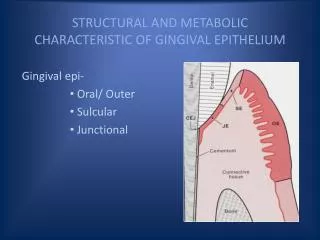
Gingival Disease. Nield-Gehrig CH 10 Perry CH 6. Gingival Description. 6 Gingival Characteristics. Color Size Position of margin Shape of margins and papillae Texture and consistency Bleeding and/or exudate. Learning to Look at the Gingiva. Healthy Gingiva.

Gingival Disease
E N D
Presentation Transcript
Gingival Disease Nield-Gehrig CH 10 Perry CH 6
6 Gingival Characteristics • Color • Size • Position of margin • Shape of margins and papillae • Texture and consistency • Bleeding and/or exudate
Healthy Gingiva • Tissue fits snugly around the tooth • Pointed papillae fill embrasure spaces • Firm and resilient • Little or no gingival crevicular fluid (GCF)
Gingivitis • The mildest and most common form of periodontal disese • Gingivitis – inflammation of the gingiva causing tissue to become edematous and erythematous…bleeds easily on provocation.
Tissue Color In Gingivitis • Acute inflammation = increased blood flow = RED tissue • Chronic inflammation = bluish-red or purplish – red
Gingival Bleeding on Probing • The two earliest signs of ginigval inflammation preceding established gingivitis are: • 1. Increased gingival crevicular fluid production rate • GCF = inflammatory exudate • Recently – development of tests for the detection or prediction of periodontal disease using the components, origin, and function of GCF • Drugs in GCF – tetracycline and Metronidazole • 2. Bleeding from the gingivl sulcus on gentle probing
Gingival Bleeding • In gingival inflammation, histopathologic alterations that result in abnormal gingival bleeding include dilation and engorgement of the capillaries and thinning or laceration of the sulcular epithelium. • Because the capillaries are engorged and closer to the surface, and the thinned, degenerated epithelium is less protective, stimuli that are normally innocuous cause rupture of the capillaries and gingival bleeding. • The severity of the bleeding and the ease of its provocation depend on the intensity of the inflammation. • In cases of moderate or advanced periodontitis, the presence of bleeding on probing is considered a sign of active tissue destruction.
Gingival Bleeding Associated with Systemc Changes • In some systemic disorders, gingival hemorrhage occurs spontaneously or after irritation and is excessive and difficult to control. • Vascular abnormalities – Vit. C deficiency or allergy • Platelet disorders – thrombocytopenic purpura • Hypprothrombinemia – Vit. K deficiency • Other coagulation defects – hemophilia, leukemia, • Deficient platlet thromboplastic factor (PF3) resulting from uremia, multiple myeloma and ostrubella purpura.
Gingival Bleeding Associated with Systemc Changes • Hormonal replacement therapy • Oral contraceptives • Pregnancy • Menstral cycle • Diabetes • Medications: • Anticonvulsants • Antihypertensive calcium channel blockers • Immunosuppressant drugs • aspirin
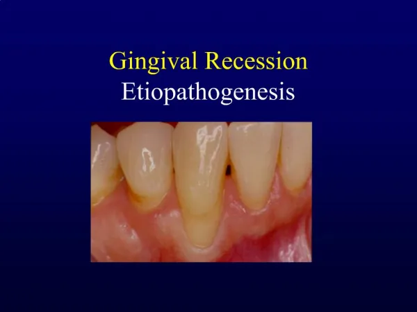
Tissue Size in Gingivitis • Increase in tissue fluid causes enlargement of the marginal and interproximal gingival tissues (Edema) • Change can be localized to a few areas or affect the whole mouth (generalized)
Changes in the Consistency In Gingival Disease • Both chronic and acute inflammations produce changes int the normal firm and resilient consistency of the gingiva. • Chronic gingivitis = edematous – (destructive) and fibrotic – (repairative) changes coexist • The consistency of the gingiva is determined by their relative pedominance.
Surface Texture Changes in Disease • The surface of normal gingiva usually exhibits numerous small depressions and elevations = stippling • In chronic inflammation the surface is either smooth and shiny or firm and nodular • This depends on whether the dominant changes are exudative or fibrotic
Smooth, Shiny Tissue • Can be: • exudative • Epithelial atrophy in atrophic gingivitis • Chronic desquamative gingivitis can also have peeling of the surface
Changes in surface texture chronic gingivitis • Hyperkeratosis results in a leathery texture (example = chronic gingival disease in a smoker) • Fibrotic = firm nodular • Drug induced gingival overgrowth also produces a nodular surface
Assess the Following • Color • Size • Position of gingival margin • Shape of margins and papillae • Use air and probe to determine texture • Consistency • Check for bleeding
More on Size • Gingival Enlargement or gingival overgrowth are the current terms used to describe an increase in the size of the gingiva • “hypertrophic gingivitis” or “gingival hyperplasia” may have erroneous pathologic connotations • Gingival enlargement is a purely clinical term
Gingival enlargement can beclassified according to etiologic factors and pathologic changes • I. Inflammatory enlargement • A. Chronic • B. Acute • II. Drug-induced enlargement • III. Enlagements associated with systemic diseases or conditions • IV. Neoplastic enlargement (gingival tumors) • V. False enlargement
Criteria of location • Localized: limited to the gingiva adjacent to a sengle tooth or group of teeth. • Generalized: involving the gingiva throughout the mouth • Marginal: confined to the marginal gingiva • Papillary: Confined to the interdental papillae • Diffuse: Involving the marginal and attached gingivae and papillae. • Discrete: An isolated sessile or pedunculated, tumorlike enlargement.
Non-Plaque –Induced Gingival diseases • Bacterial origin – • Neisseria gonorrhea – associated lesions • Treponema pallidum – associated lesions • Streptococcal species – associated lesions • RARE – Non-plaque is RARE
Non-Plaque-induced gingival diseases of viral origins • RARE – Non-plaque is RARE • Acute herpetic gingivostomatitis • Recurrent oral herpes • Varicella-zoster infections
Primary Herpetic Gingivostomatitis • Caused by herpes simplex virus type 1 (HSV-1) • Most often occuring in infants and children under 6 years • In most people the primary infection is asymptomatic • As part of the primary infection, the virus ascends through sensory and autonomic nerves, where it persists as latent HSV in neuronal ganglia that innervate the site • In 1/3 of the world’s population secondary manifestations result from various stimuli
NUG • Punched-out, craterlike depressions at the crest of the interdental papillae • Gray, pseudomembranous slough • Linear erythema • Spontanious gingival hemorrhage • Fetid odor
Gingival Diseases Modified by Malnutrition • Most clinical studies have not shown a relationship between the development of gingival diseases and malnutrition with the possible exception of severe vitamin C deficiency.
Lichen Planus • Inflammatory mucocutaneous disorder that may involve mucosal surfaces and the skin. • Current evidence suggests that lichen planus is an immunologically mediated mucocutaneous disorder in which host T lymphocytes play a central role.