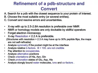
Refinement of a pdb-structure and Convert
100 likes | 349 Vues
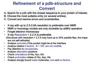
Refinement of a pdb-structure and Convert. A. Search for a pdb with the closest sequence to your protein of interest. B. Choose the most suitable entry (or several entries). C. Convert and resolve errors and uncertainties. X-ray with up to 2.5-2.8A resolution is preferable over NMR

Refinement of a pdb-structure and Convert
E N D
Presentation Transcript
Refinement of a pdb-structure and Convert • A. Search for a pdb with the closest sequence to your protein of interest. • B. Choose the most suitable entry (or several entries). • C. Convert and resolve errors and uncertainties. • X-ray with up to 2.5-2.8A resolution is preferable over NMR • NMR or homology models are only dockable by skillful operators • Forget electron microscopy • X-ray Resolution < 2.2 A is preferable. • (Structures with resolution > 2.3 A may have up to 30% peptide flips, the maps are not self-refinable) • Analyze symmetry if the pocket might be at the interface • Analyze relative b-factors. B > 100. are not credible • Pay attention to occupancies. • Analyze alternative positions • Check orientations of His, Asn, Gln • Check protonation states of Glu, Asp, His • Analyze stongly bound water molecules, ions and co-factors .
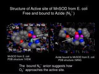
Preparations: symmetry Symmetry related subunit Problem: the true pocket is formed by chains which are not explicitly present in a pdb entry. Goal: Find all molecules/subunits or chains involved in the interaction with the ligand. Warning signs: ICM pocket finder does not show pocket density; Binding site is obviously exposed Recovery: generate symmetry related subunits (View/Cryst.Cell) Example: Cycloldextrin glycosyltransferase Entry: 1cdg, Res. 2.0A (Docking Rmsd without symmetry: 9.76) More examples: transthyretin 1f41 (thyroid hormone binds at the dimer interface)
Preparations: occupancies, b-factors and alternatives Glossary: B-factor (or temperature factor): mean-square displacement of atom from its position in the model. Bi = 79*<u2> (B of 80 means 1A dev.) Normal range: 5. – 50. A2. Occupancy: A fraction of atomic density at a given center. It there are two equally occupied conformers, both will have occupancies of 0.5 Normal value: 1. Range: 0.-1. Alternatives: If two or more alternative conformations for the same atom or group are discernable in the density, several alternative sets of coordinates are deposited. Occupancies <= 0.5 are shown in magenta High b-factors are colored red Problem: sometimes, when electron density is poor and/or ambiguous, crystallographers make things up (or just deposit an arbitrary conformation from a refinement program) Goal: Identify fantasy atoms/groups Warning signs: occupancies less than 0.5, b-factors larger than 60-80 A2. Tool: Color/label pocket atoms by occupancies/b-factors. Recovery: Choose another entry, or refine with a ligand, or perform restrained minimization. Choose one of alternatives, or create alternative models
Preparations: occupancies, b-factors and alternatives. Example. This is a very high resolution structure. For some key residues two alternative conformations are provided. Recovery: Choose one alternative or generate several separate docking models Alternative positions for Thr and Val32 Entry: 1hmt. Res. 1.4 Fatty Acid Binding Protein with stearic acid
Preparations: fixing histidines Orientation at the heavy atom level We need to discriminate between These two conformations Often the xi2 angle needs to be Corrected by 180 degrees. That is how histidine density really looks Uncertainly at the protonation level You need to decide which of the three conformations is correct for each important location. The charged conformation is rare. + d d e e Problem: orientations and protonation states of histidines are frequently wrong on pdb entries and need to be fixed to ensure correct docking results. Placement principle: maximization of hydrogen bonds and other interactions with the rest of the protein and/or with the ligand. Recovery: ICM procedure optimizeHisProAsnGln finds the best orientation and protonation state
Preparations: determining orientations of Gln, Asn, side chains Orientation at the heavy atom level The two conformations shown give similar electron density. We need to discriminate between these two conformations of the Asn side chains. The same ambiguity needs to be resolved for the xi3 angle of Gln Background: xi2 in asparagines and xi3 in glutamines are frequently wrong or undefined and need to be corrected ensure correct docking. Placement principle: maximization of hydrogen bonds and other interactions with the rest of the protein and/or with the ligand. Recovery: ICM optimizeHisProAsnGln procedure.
Preparations: do I need to uncharge Asp, Glu, Lys and Arg? Definitions: DERK is Asp (D), Glu (E), Arg(R) or Lys (K) Facts: pKs: His 6.0, Cys 8.3 Glu 4.2, Asp 3.9 General recommendation: keep the DERK residues charged. Problem: while in most cases DERKs are charged, in some special cases ED need to be uncharged or His needs to be charged. Warning signs: a DERK is buried and NOT involved in a salt bridge; Several DERKs of the same kind/charge are pointing to the same space. Example: HIV protease. 1ida. Asp 25 and 25’ are protonated. Recovery: Modify them to the uncharged forms.
Preparations: which waters to keep? Example: 1eye dihydropteroate synthase, anti-mycobacterial/TB target. It binds to the buried Asp177 and improves electrostatic desolvation by ~10 units. Definition: crystallographic water: an oxygen placed by a crystallographer or a refinement program to a blob of electron density. General recommendation: get rid of all water molecules, Keep only water molecules with three or four hydrogen bonds with the protein or ligand atoms. Reason: keeping inappropriate water(s) will prevect correct docking, while dropping good waters is usually tolerated. Howeversome tightly bound water molecules help docking and scoring and prevet from erroneous placement of H-bond-rich ligand groups in water sites. Recovery: Find interface waters with 3 or more protein/ligand neighbors and include them into your model.
Preparations: cofactors and metals? Problem: metals may be required to dock a charged native ligand (e.g. ATP is charged and requires 2 Mn++ ions.) However, to the metals may not always be necessary for docking of neutral drugs. Example: a kinase domain. 1atp
Local quality: Energy Strain • More sensitive that geometrical and clash criteria • Based on fast ICM calculation of residue energies (recently accelerated 1000 fold) • Energy = Vacuum Energy + Solvation + Entropy • Deriving energy distributions for each amino-acid type • All high resolution PDB structures (<1.5A) collected • Distributions of residue energies calculated • Energy Distribution for each amino acid derived • Normalized energies derived Calculating normalized residue energies for a model • Calculate Z-score (normalized energy) for each residue • Residues with Enorm > 5 are probably wrong Maiorov, Abagyan, 1998 Proteins, The fast version: 2004