Crystallography: Structure Solution and Basic Refinement
Learn about crystallography, experiment files, structure solution methods like direct and Patterson methods, and software recommendations.


Crystallography: Structure Solution and Basic Refinement
E N D
Presentation Transcript
Recommended software • Shelx • http://shelx.uni-ac.gwdg.de/SHELX/ • WinGX • http://www.chem.gla.ac.uk/~louis/software/wingx/ • Platon • http://www.chem.gla.ac.uk/~louis/software/platon/ • Solution programs (see later) • A good text editor • e.g. Notepad++ • http://notepad-plus.sourceforge.net/uk/site.htm
Chemistry Facilities • Bruker Smart Apex CCD Diffractometer
From crystal to cif • Get crystal • Short experiment to index • Long experiment to collect complete data • Integration • Absorption correction • Identify space group • Solve You start here • Refine • Cif
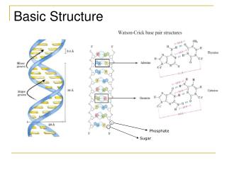
What is crystallography all about • A crystal is a 3D periodic array of molecules • X-rays interact with (diffract from) electrons • Diffraction results in a regular pattern of spots (due to constructive and destructive interference) • Intensities observed are related to atom types and positions • We build a model and compare the calculated diffraction pattern with the observed
What you will get from us • 2 or 3 files • ins file • The shelx instruction file contains unit cell, radiation wavelength, temperature, crystal system and space group information, unit cell contents from user input • hkl file • The reflection data, contains indexed intensities with associated estimated standard deviations • cif file • Contains some experimental details (not essential)
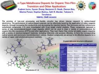
Files – ins file TITL pg24 in P-1 CELL 0.71073 10.724 12.440 12.643 72.40 79.36 73.33 ZERR 2.00 0.004 0.005 0.005 0.02 0.02 0.02 LATT 1 SFAC C H N S HG UNIT 52 106 2 6 2 PATT HKLF 4 END
Files – hkl file -12 0 1 64.46 2.70 -12 -1 2 47.71 3.70 -12 -1 1 41.90 3.48 -12 -2 1 15.71 0.80 -12 -2 2 41.02 1.56 -12 -3 2 20.34 0.97 -12 -3 1 2.96 0.62 -12 -4 1 27.33 1.27 -12 -4 2 21.33 1.05 -12 -5 1 60.67 2.48 -12 -6 1 66.61 2.61 -11 -8 2 9.18 0.73 -11 -8 1 37.45 1.50 -11 -7 1 3.94 0.59 -11 -7 2 92.96 4.46 -11 -7 3 46.55 2.36 -11 -6 3 16.54 0.86 -11 -6 2 135.97 3.44 -11 -6 1 9.06 0.72
Structure Solution • We need to get an initial model from which to work • This is called structure solution • We can only measure intensity but we really want to know the phase of each reflection. • Several methods of extracting initial estimated phases from our data are available.
Structure solution • Direct methods • Requirements • It is desirable but not required to have a centrosymmetric structure. • How it works • Uses statistical relationships between the intensity of different reflections to establish phases. • Look out for… • This is a very powerful method which often works very well but as it is based on statistical analysis, it will sometimes fail. • Computationally demanding. Scales inversely with symmetry, i.e. low symmetry large unit cells take more time.
Structure solution • Direct Methods Programs • xs from the shelx suite (works for me 99% of the time) • Instruction is TREF • You may specify a number after TREF to increase the number of trials for difficult structures. • Sir software • Several versions • Sir92, Sir97, Sir2002, Sir2004 • May get different results with different versions • Variety of options for structures of different size/difficulty OR • Can be set up through, e.g. WinGX for simplicity but less control.
Structure solution • Patterson Methods • Requirements • A heavy element, e.g. Fe, Cl, S, etc • How it works • Generates a map of ‘peaks’ representing difference vectors, i.e. interatomic vectors • Peak intensity is related to the product of the atomic numbers of the two atoms involved thus the heaviest elements are identifiable. • Look out for… • Depending on the program you may only get the heavy atom positions. Completing the structure may take more time and effort than e.g. Direct methods
Structure Solution • Patterson Methods Programs • xs from the shelx suite • Instruction is PATT • You will only get heavy atom positions back • Dirdif • Will attempt to complete the model and guess atomic assignments. • Can be extremely useful for complicated metal clusters, etc.
Structure Solution • Partial Structure Expansion • Requirements • Knowledge of expected substructures and their geometries, e.g. a benzene ring • How it works • Uses a Patterson map and rotates the substructure around three axes until a best fit is found to the data. There will then be attempts to complete the model using the phase information from your partial structure as a basis for phase refinement. • Look out for • Not many pitfalls assuming you are confident of the unit cell contents. • More time consuming to set up than other methods and unsuitable for unknown samples.
Structure Solution • Partial structure expansion programs • PATSEE from the wingx suite • You must provide a list of coordinates for your substructure. • Dirdif • Provides a small database of common geometries e.g. benzene rings, indoles or can use your own. • Not necessarily user friendly in my opinion
Structure solution • Charge flipping • Requirements • Complete data (at the moment) • How it works • Use random phases, ‘flip’ the sign of electron density charge below a threshold and get new phases. Use new phases with original magnitudes. Repeat. • No symmetry is used for solution. It is determined afterwards. • Look out for… • Unreliable results with incomplete data
Structure solution • Charge-flipping software • Superflip • From originators of the method • Available as standalone (not recommended), via WinGX, via crystals • Flipper • Available in Platon
Structure solution • Has it solved? • Use your chemical knowledge… • How atoms interact • E.g. expected bond distances and angles for particular arrangements • Reactions or decompositions which may occur • Non-bonded interaction lengths and types • E.g. +ve to +ve is not going to be common • Charge • Should always be neutral for a unit cell • Assignment of charge on metals, ligands • Likely patterns of motion • E.g. neighbouring atoms are likely to have similar thermal motion • Spinning or wagging of certain groups may be expected • Groups which are likely to be rigid, move as one
Structure solution • Watch out for… • If provided, don’t assume atomic assignments are correct. • Look for expected geometry, e.g. rings, octahedra • Might get messy q-peaks and need to trim back to your structure. • Don’t assume that if you can’t see your compound that is isn’t there and just obscured by noisy peaks • Incorrect atomic assignment and missing atoms will affect the calculated phases and may mean some atoms don’t appear at first. • Be patient • Try any and all structure solution programs you can find • Different programs produce different results even using the same method. • Those mentioned are not the only ones but should be sufficient.
Structure Solution example • Data taken from Oxford primer: • Crystal structure determination • William Clegg
Refinement • Iterative process • Be patient • Sometimes it takes a long time and is difficult • Sometimes it is easy and quick • You must model all electron density (q-peaks) or be able to explain why modelling some peaks is not appropriate. • Structure must be chemically reasonable • Pay attention to • Geometry • R-factors • Q-peaks • ADP’s • Contacts
Typical Refinement START Switch current atoms to anisotropic converged Yes No All atoms anisotropic? Weighting scheme FINISH! Initial solution not converged None All atoms correctly identified? Yes Yes Refine… Problems? yes Model complete? Large Q-Peaks Missed Atoms? yes No No Odd sized or Shaped ADP’s Disorder? Fourier difference map Check atomic assignments Switch unusual atom(s) to isotropic refinement Wrong atom type(s)?
Refinement • Common problems • Wrongly assigned atoms • Disorder, particularly solvent* • Twinning* • Incorrect space group • Q-Peaks due to strong absorption (heavy metals present) • Not all refinements will end happily • You may have to leave some atoms isotropic • You may be unable to find or place hydrogen atoms • You may have a high R-factor • You may simply not be able to finish due to the above issues or poor quality data * Workshops on these will be given later
TITL SAMPLE in Pbca CELL 0.71073 10.7308 16.4002 18.9778 90.000 90.000 90.000 ZERR 8.00 0.0005 0.0007 0.0008 0.000 0.000 0.000 LATT 1 SYMM 0.5-X, -Y, 0.5+Z SYMM -X, 0.5+Y, 0.5-Z SYMM 0.5+X, 0.5-Y, -Z SFAC C H N O UNIT 160 152 8 32 TEMP -123 L.S. 4 BOND $H FMAP 2 ACTA CONF PLAN 20 WGHT 0.040400 1.691300 FVAR 0.13360 O1 4 1.183095 0.526967 0.621173 11.00000 0.04325 0.03499 = 0.02675 -0.00293 -0.00093 -0.01115 C2 1 1.183370 0.488078 0.567136 11.00000 0.02854 0.02571 = 0.02494 0.00436 -0.00205 -0.00215 O3 4 1.266176 0.500831 0.515249 11.00000 0.03273 C4 1 1.239322 0.447764 0.456425 11.00000 0.03222 HKLF 4 REM HL9005 in Pbca REM R1 = 0.0452 for 3267 Fo > 4sig(Fo) and 0.0476 for all 3421 data REM 226 parameters refined using 0 restraints END WGHT 0.0404 1.6913 REM Highest difference peak 0.267, deepest hole -0.198, 1-sigma level 0.039 Q1 1 0.8842 0.3387 0.6174 11.00000 0.05 0.27 Q2 1 0.9964 0.3517 0.6038 11.00000 0.05 0.24 Q3 1 0.9221 0.3187 0.5592 11.00000 0.05 0.23 Q4 1 0.6446 0.2208 0.7061 11.00000 0.05 0.22 Q5 1 0.9203 0.3060 0.4928 11.00000 0.05 0.21 Q6 1 0.9370 0.3938 0.5769 11.00000 0.05 0.20 Shelx • Anatomy of a shelx file
Restraints and constraints • A restraint allows a parameter to refine within limits • E.g. like applying a spring • A constraint fixes a parameter. It is not allowed to refine. • E.g. like applying rope • Restraints are treated as additional observations • Restraints have an associated e.s.d. i.e. a measure of how strict the restraint should be • Most commands have reasonable defaults • Use to correct poor geometry with chemical knowledge • Can use temporarily to maintain reasonable geometry when identifying a problem, e.g. disorder • Only use when necessary ensure e.g. distance restraints use an appropriate value, e.g. taken from CSD data • Make sure there isn’t an underlying problem before resorting to R&C • More on these in disorder workshop.
Atomic assignment problems • Models are a picture of electron density • Different elements have different numbers of electrons • Incorrect assignments should show up in thermal parameters. • Too small an element will cause the ADP to shrink • Too large an element will cause it to grow • Why?
Atom is actually a nitrogen which has higher z and therefore e- density than our modelled carbon. Therefore the carbon ‘shrinks’ to increase its e- density Atom is actually a carbon which has lower e- density than our model. Therefore the nitrogen ‘grows’ to spread out its e- density Atomic assignment problems
Hydrogen atoms • Difficult to find • Only one electron to diffract from • Heavier elements further obscure hydrogen positions • Incorrectly located • Diffract from electrons not nuclei!!! • Valence electron only, located ‘in bond’
Hydrogen placement • Find them if you can • They may often be visible in a difference map • You may then be able to refine, partially refine them or constrain them. • Geometric placement • Uses expected (e.g. tetrahedral) angles and distances appropriate for x-ray diffraction • HFIX mn • m specifies how to place e.g. tetrahedral angles • n specifies how to refine, e.g. full coords or riding
Hydrogen Placement • Common HFIX types 1 2 3 4 8
Some Quality indicators • R1 • The conventional R-factor • <10% is publishable • <5% is good • Goof • Goodness of fit • Should be as close to 1 as possible • More than around 0.4 away is cause for concern • Rint • An R-factor for data merging of equivalent reflections • If perfect would be 0 • A rough guide is to expect R1 to be close to this value • Max Shift • The max shift of all parameters from non-linear least squares refinement • Should be zero for a properly converged, finished model.
Finishing off • Confident the structure is ok? • Platon checkcif • IUCr web tool • http://checkcif.iucr.org/ • Platon • Does additional checks if FCF file is present • Make sure it is up to date!