Achilles Ruptures
Achilles Ruptures. Michael Lapner 2009. Case History. 65 y.o . female in ER, felt pop in ankle, mild pain Thompson + ER doc wants to know what you would like to do?. Physical Exam. Imaging. Surgery. Post-Op Protocol?. Objectives. Epideomiology Anatomy History Exam Findings

Achilles Ruptures
E N D
Presentation Transcript
Achilles Ruptures Michael Lapner 2009
Case History • 65 y.o. female • in ER, felt pop in ankle, mild pain • Thompson + • ER doc wants to know what you would like to do?
Objectives • Epideomiology • Anatomy • History • Exam Findings • Surgery vs Non-Operative treatment? • Percutaneousvs Open? • Chronic Rupture management • Post-Op Protocol • Summary
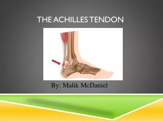
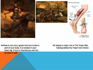
Largest most powerful tendon in ankle • Two muscle units • Gastrocnemius & soleus • Origins • Gastrocnemius • Strongest in plantar flexion, knee extended • Soleus • Strongest in plantar flexion, knee flexed
Anatomy • Complex rotates 90 degrees toward insertion to calcanealtuberosity. • Gastrocfibres end lateral • soleus medial • Can think of this as internal rotation
Vascularity • Through paratenon on deep surface • Muscle arterial branches from G-S complex proximally • Intraosseus vessel at insertion of tendon into calcaneus. • Avascular zone 2-6 cm proximal from insertion
Risk Factors • The weekend warrior • Obesity (both men and women) • HTN • DM association (< 44 y.o.) • Fluoroquinolones • Steroids • Previous history rupture • infection
Definitions • Tendinopathy • Tendinitis • Inflammation • Tendinosis • Small tears in connective tissue
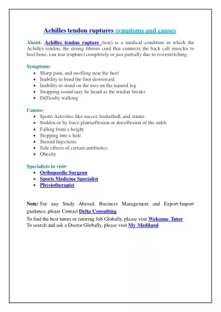
History • Typically 30-50 y.o. male • Previous healthy • No prior leg injuries • Left foot more common • Up to 25% missed in ER (edema)
History • Sudden snap • Acute severe pain (not always) • Limp • Complain swelling • Recent increase physical activity
Epidemiology • 30-50 • training, intensity, activity change • New activity • Almost every sporting activity • Running sports
Cause • Sudden forced plantar flexion • Unexpected dorsiflexion • Violent dorsiflexion of plantar flexed foot • Direct injury • Laceration, blow to posterior leg
Physical Exam • Examine entire length of complex • Tenderness, swelling, ecchymosis, defects • Gapping • Unable to stand on toes
Special Tests • Hyperdorsiflexion sign • Prone, knees 90, maximal DF increased • Thompson Test • Prone, squeeze calf • O’Brien Test • Needle 10 cm proximal to calcaneal insertion • Tilts rostral when intact.
Differential • Fracture • Sprain • CFL injury • TFL injury
Differential • Achilles Tendinosis • Bony injury • Fascial tear • Inflammatory • Syndesmosis • Tennis leg (tear plantaris) • Vascular injury
Radiograph • In order to rule out other injuries • Soft tissue swelling • Accessory ossicles • Fractures • Haglund deformity
Ultrasound • Thickness • Character • Presence of tear • MRI • Paratenonitis, tendinosis, bursitis
Treatment • Surgical versus non-operative? • Open versus percutaneous?
Non-operative • Serial casting • Plantar flexed position • Progression over time to plantigrade • Boot walker/heel insert. • Early/functional protocol – no definition
Early/Later Non-Op comparison • 2006 study • Non wt-bearing cast immobilization versus immediate wt bearing. • Three 1.5 cm heel lifts initially • Decrease by one lift q 2 / 52 • Complication rate same • No change strength. Costa 2006, JBJS
Op/Non-Op Rx RCT • 50 patients non-surgically/open repair • Groups – equinus cast 10 days • Orthosis 20 plantar flexion • At 4 weeks brought to neutral. • 6 weeks WBAT • 8 weeks weaned • No difference in motion/calf circumference/strength/ re-rupture rate/complication at 1 year Twaddle/Poon AJSM 2007
Operative • Up to 33 % higher complication rate • Re-rupture 3x lower than non-operative • Infection (1%) • Fistulae (3%) • Rerupture (2%) • Minor complications • Skin/tendon necrosis (2%)
Percutaneous? • Lower infection rate • Higher rate of injury to sural nerve
Non-operative • Early reports suggest re-rupture > 40% • Newer protocols, shorter immobilization • Rerupture rate comparable to surgery • Nature taking it’s course?
Evidence Non-Operative • Surgical/non-op treated same rehab protocol • Achilles repair (krackow) • 10 days POP, equinus • Non-op directly casted • Cast removed, removable below knee orthosis • 20 degrees PF • 5 minutues off/hr • Sitting, leg hanging, practice A-DF/P-PF Twaddle BC, Poon P.Am J Sports Med. Dec 2007.
Evidence Non-Op • 4/52 out, orthosis – neutral • Continue ROM • 6/52 out, WBAT, wearing orthosis • Remove at night • 8/52 out, patients weaned off
Results • 3 cases re-rupture • 2 surgical • 1 fell down stairs • 1 hit by car trying to stop robbery • 1 non-operative • Slipped off embankment • All re-ruptures treated with surgery
Review of all conservative • Skin complications 0.5 % • 8.5% unspecified • Major 0.6% • Re-rupture 9.8%
Percutaneous • Ma Griffith described in 1977 • 18 tendons, stab incisions • Surgeons knots prox/distal ends, approximate • Short leg cast, 4 weekns non wt-bearing • 4 weeks wt-bearing low heel cast
Percutaneous • Later studies, minor variations • High rates of sural nerve entrapment • 16.7 % Elliot RR, Calder JD.Foot Ankle Clin. Dec 2007;12
Percutaneous safer? • Cretnik et al, Ceccarelli et al, Lansdaal et al., • AJSM, CORR, I
Open • Medial longitidunal incision • Plantaris • Avoid sural nerve • Midline • Higher rates of complications (wound) • Higher rates of adhesions
Open • Paratenon divided longitudinally • I&D ends • Reapproximated with heavy non-absorbable • Ticron • Modified kessler • Krackhow • Bunnel • Tijima
Open • Ankle in PF • Short period immobilization • Then foot brought to neutral rigid orthosis • Partial wt bearing commenced • Immobilization D/C after 4-6 weeks • Active/active assisted begun • Full activities at 4 months
Open • Young/athletic • Increased muscle strength • Power • Endurance • Earlier return to activities
Quantitave Review of Operative and Non Operative management of achilles tendon ruptures • 2002, Wong et al., • retrospective and prospective data • 125 journals • 5370 patients • Re-rupture highest in immobilized/conservative • Complications highest in percutaneous • Early mobilization best (trend) • Open, best functional results, acceptable comp.
RCT 2007 • 2 groups, open / absorbable technique • Early motion PF vs immobilized cast at neutral • Tendon elongation greater in cast immobilized at neutral • Used markers intraop /U/S to measure distraction Kangas 2007 AJSM
Use of augmentation? • 2007 study, 17 month F/U • No difference in re-rupture rate. • Increased time/morbitidy of harvesting plantaris unnecessary
DVT prophylaxis • Controversial • Lapidus et al., JOT 2007