The Skeletal and Muscular Systems
620 likes | 795 Vues
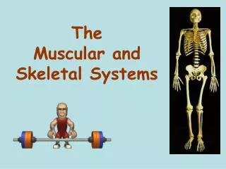
The Skeletal and Muscular Systems. By Dr Shamshad Begum Loni. LECTURE NOTES. Axial skeleton skull (cranium and facial bones) hyoid bone (anchors tongue and muscles associated with swallowing) vertebral column (vertebrae and disks) thoracic cage (ribs and sternum)

The Skeletal and Muscular Systems
E N D
Presentation Transcript
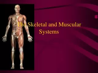
The Skeletal and Muscular Systems By Dr Shamshad Begum Loni LECTURE NOTES
Axial skeleton skull (cranium and facial bones) hyoid bone (anchors tongue and muscles associated with swallowing) vertebral column (vertebrae and disks) thoracic cage (ribs and sternum) Appendicular skeleton pectoral girdle (clavicles and scapulae) upper limbs (arms) pelvic girdle (coxal bones, sacrum, coccyx) lower limbs (legs)
The Human Skeleton 1. Carpals 2. Cranium 3. Femur 4. Innominate 5. Mandible 6. Metacarpals 7. Metatarsals 8. Phalanges 9. Rib 10. Scapula 11. Sacrum 12. Vertebra 1. Clavicle 2. Fibula 3. Humerus 4. Patella 5. Radius 6. Sternum 7. Tarsals 8. Tibia 9. Ulna
What are the Five main functions of the human skeleton • Protect the vital organs we talked about last week • 2. Give us shape • 3. Allow us to move because our muscles • are attached to our bones • 4. Storage of nutrients such as calcium and silicon • 5. Formation of blood cells
Types of Bone Cells • Osteocytes Mature bone • Osteoblasts(remove calcium from blood and build • new matrix. They become trapped • osteoclasts) • Bone-forming cells • Osteoclastsremove damaged cells and release • calcium into blood (Bone-destroying cells) • Break down bone matrix for remodeling and release of calcium • Bone remodeling is a process by both osteoblasts and osteoclasts
Interesting Facts about theSkeletal System • Do we have more bones when we are a baby or when we are all grown up? • Baby has 305 bones and an adult has 206 bones. This is because as we grown some of our bones join together to form one bone. • The longest bone in our bodies is the femur (thigh bone). • The smallest bone is the stirrup bone inside the ear. • Each hand has 26 bones in it. • our nose and ears are not made of bone; they are made of cartilage, a flexible substance that is not as hard as bone. Compact bone osteocytes within lacunae arranged in concentric circles called lamellae This surround a central canal; complex is called Haversian system Canaliculi connect osteocytes to central canal and to each other
Structures Associated with the Synovial Joint • Bursae – flattened fibrous sacs • Lined with synovial membranes • Filled with synovial fluid • Not actually part of the joint • Tendon sheath • Elongated bursa that wraps around a tendon
Types of Joints Hinge- A hinge joint allows extension and retraction of an appendage. (Elbow, Knee)
Types of freely movable joints Saddle: carpal and metacarpal bones of thumb Ball and socket: shoulder and hip joints Pivot- rotation only: proximal end of radius and ulna Hinge- up and own movement in one plane: knee and elbow Gliding- sliding and twisting: wrist and ankle Condyloid- movement in different planes but not rotations: btw metacarpals and phalanges
Ball and Socket- A ball and socket joint allows for radial movement in almost any direction. They are found inthe hips and shoulders. (Hip, Shoulder)
Gliding- In a gliding or plane joint bones slide past each other. Mid-carpal and mid-tarsal joints are gliding joints. (Hands, Feet)
Saddle- This type of joint occurs when the touching surfaces of two bones have both concave and convex regions with the shapes of the two bones complementing one other and allowing a wide range of movement. (Thumb)
Types of movement and examples (with muscles) flexion- move lower leg toward upper extension- straightening the leg abduction- moving leg away from body adduction- movong leg toward the body rotation- around its axis supination- rotation of arm to palm-up position pronation- palm down circumduction- swinging arms in circles inversion- turning foot so sole is inward eversion- sole is out
Elevation and depression- raising body part up or down Aging and bones both bone and cartilage tend to deteriorate cartilage: chondrocytes die, cartilage becomes calcified osteoporosis bone is broken down faster than it can be built bones get weak and brittle; tend to fracture easily Risk factors for osteoporosis Inadequate calcium Little weight-bearing exercise Drinking alcohol, smoking Being female: decreased estrogen secretion after menopause
Types of bone breaks Simple- skin is not pierced Compound- skin is pierced Complete- bone is broken in half Partial- broken lengthwise but not into two parts Greenstick- incomplete break on outer arc Comminuted- broken into several pieces Spiral- twisted
Osteoporosis • Osteoporosis is a term that means "porous bones.”. • Osteoporosis is a condition in which bones have lost mineralsespecially calcium, making them weaker, more brittle, and susceptible to fractures (broken bones)., • the most common places where fractures occur are the back (spine), hips, and wrists.
Scurvy • We depends on exogenous dietary sources to meet vitamin C needs. • Consumption of fruits and vegetables or diets fortified with vitamin C are essential to avoid ascorbic acid deficiency. • Even though scurvy is uncommon, it still occurs and can affect adults and children who have chronic dietary vitamin C deficiency.
Bursitis • Inflammation of the Bursa (fluid filled sac surrounding the joint). • A bursa can become inflamed from injury, infection (rare in the shoulder), or due to an underlying rheumatic condition. • Bursitis is typically identified by localized pain or swelling, tenderness, and pain with motion of the tissues in the affected area.
Skeleton and other systems Skin makes vitamin D which enhances calcium absorption Skeleton stores calcium for muscle contraction, nervous stimulation, blood clot formation Red marrow- site of blood cell formation Calcium levels regulated by parathyroid hormone and calcitonin kidneys (can help provide vitamin D) digestive system (can release calcium into blood
Muscular System Functions • Body movement (Locomotion) • Maintenance of posture • Respiration • Diaphragm and intercostal contractions • Communication (Verbal and Facial) • Constriction of organs and vessels • Peristalsis of intestinal tract • Vasoconstriction of b.v. and other structures (pupils) • Heart beat • Production of body heat (Thermogenesis)
Properties of Muscle • Excitability: capacity of muscle to respond to a stimulus • Contractility: ability of a muscle to shorten and generate pulling force • Extensibility: muscle can be stretched back to its original length • Elasticity: ability of muscle to recoil to original resting length after stretched
Interesting facts about the Muscular System • Muscle: A tissue composed of fibers capable of contracting to effect bodily movement • There are about 650 muscles in the human body.
Some Muscles 1. Gastrocnemius 2. Sartorius 3. Deltoid 4. Sternocleidomastoid 5. Tibialis 6. Hamstring group 7. Rectus Abdominus 8. Triceps 9. Biceps 10. Extensor Group
Types of muscles Skeletal muscles: Attached to bones. (what happens when you extend your arm?) Smooth muscle: Surround organs, tubes, eg. stomach, urinary bladder, blood vessels. Contract propels content through organs (eg. expel urine). Cardiac muscles: Heart muscle makes your heart pump blood. Tendons Muscle Blood vessel Connective tissue Muscle fibre
Muscle Classification Functionally 1. Voluntarily 2. Involuntarily Structurally 1. Striated 2. Smooth Combined 1. Visceral 2. Cardiac 3. Skeletal
Nerve and Blood Vessel Supply • Motor neurons • stimulate muscle fibers to contract • Neuron axons branch so that each muscle fiber (muscle cell) is innervated • Form a neuromuscular junction (= myoneural junction) • Capillary beds surround muscle fibers • Muscles require large amts of energy • Extensive vascular network delivers necessary oxygen and nutrients and carries away metabolic waste produced by muscle fibers
Basic Features of a Skeletal Muscle • Muscle attachments • Most skeletal muscles run from one bone to another • One bone will move – other bone remains fixed • Origin – less movable attach- ment • Insertion – more movable attach- ment
Basic Features of a Skeletal Muscle • Muscle attachments (continued) • Muscles attach to origins and insertions by connective tissue • Fleshy attachments – connective tissue fibers are short • Indirect attachments – connective tissue forms a tendon or aponeurosis • Bone markings present where tendons meet bones • Tubercles, trochanters, and crests
Skeletal Muscle Structure • Composed of muscle cells (fibers), connective tissue, blood vessels, nerves • Fibers are long, cylindrical, and multinucleated • Tend to be smaller diameter in small muscles and larger in large muscles. 1 mm- 4 cm in length • Develop from myoblasts; numbers remain constant • Striated appearance • Nuclei are peripherally located
Muscle Fiber Anatomy • Sarcolemma - cell membrane • Surrounds the sarcoplasm(cytoplasm of fiber) • Contains many of the same organelles seen in other cells • An abundance of the oxygen-binding protein myoglobin • Punctuated by openings called the transverse tubules (T-tubules) • Narrow tubes that extend into the sarcoplasm at right angles to the surface • Filled with extracellular fluid • Myofibrils -cylindrical structures within muscle fiber • Are bundles of protein filaments (=myofilaments) • Two types of myofilaments • Actin filaments (thin filaments) • Myosin filaments (thick filaments) • At each end of the fiber, myofibrils are anchored to the inner surface of the sarcolemma • When myofibril shortens, muscle shortens (contracts)
Sarcoplasmic Reticulum (SR) • SR is an elaborate, smooth endoplasmic reticulum • runs longitudinally and surrounds each myofibril • Form chambers called terminal cisternae on either side of the T-tubules • A single T-tubule and the 2 terminal cisternae form a triad • SR stores Ca++ when muscle not contracting • When stimulated, calcium released into sarcoplasm • SR membrane has Ca++ pumps that function to pump Ca++ out of the sarcoplasm back into the SR after contraction
Sarcomeres: Z Disk to Z Disk • Sarcomere - repeating functional units of a myofibril • About 10,000 sarcomeres per myofibril, end to end • Each is about 2 µm long • Differences in size, density, and distribution of thick and thin filaments gives the muscle fiber a banded or striated appearance. • A bands: a dark band; full length of thick (myosin) filament • M line - protein to which myosins attach • H zone - thick but NO thin filaments • I bands: a light band; from Z disks to ends of thick filaments • Thin but NO thick filaments • Extends from A band of one sarcomere to A band of the next sarcomere • Z disk: filamentous network of protein. Serves as attachment for actin myofilaments • Titin filaments: elastic chains of amino acids; keep thick and thin filaments in proper alignment
Sarcomere Z A Z A Z A (I) I Z Z Z
Myofilaments 1. Myosin: 110Å thick; confined to the A-band. (Mole. wt. 500,000 deltons; 200 molecules/myofilament) A. Tail- 800Å long, composed of a double helix B. Head (cross bridges)-600Å terminating in a globular double structure. Contains binding sites for actin & ATP
Myofilaments 2. Actin: 60A thick; runs from Z-line (disc) to just inside A-band. Mole wt. 60,000 deltons. G-actin (globular units): contracted form F-actin (fibrous polymers): relaxed form Actin associated proteins A. Tropomyosin B. Troponin
Neuromuscular Junction • Region where the motor neuron stimulates the muscle fiber • The neuromuscular junction is formed by : 1. End of motor neuron axon (axon terminal) • Terminals have small membranous sacs (synaptic vesicles) that contain the neurotransmitter acetylcholine(ACh) 2. The motor end plate of a muscle • A specific part of the sarcolemma that contains ACh receptors • Though exceedingly close, axonal ends and muscle fibers are always separated by a space called the synaptic cleft