Download
1 / 33
770 likes | 5.11k Vues
Trace the pathway of blood ( )through the body using the following terms: Aorta Right atrium Left atrium Right ventricle Left ventricle Lungs Vena Cava Venules Arterioles Capillaries Body tissues Pulmonary artery Pulmonary vein. The Heart. Functions of the Heart.

E N D
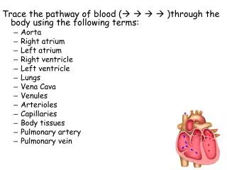
Trace the pathway of blood ( )through the body using the following terms: • Aorta • Right atrium • Left atrium • Right ventricle • Left ventricle • Lungs • Vena Cava • Venules • Arterioles • Capillaries • Body tissues • Pulmonary artery • Pulmonary vein
Functions of the Heart • Generating blood pressure • Routing blood • Heart separates pulmonary and systemic circulations • Ensuring one-way blood flow • Heart valves ensure one-way flow • Regulating blood supply • Changes in contraction rate and force match blood delivery to changing metabolic needs
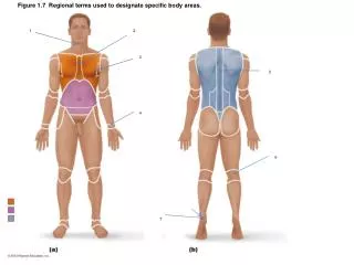
Size, Shape, Location of Heart • Size of a closed fist • Shape • Apex: Blunt rounded point of cone • Base: Flat part at opposite of end of cone • Located in thoracic cavity in mediastinum
External Anatomy • Four chambers • 2 atria • 2 ventricles • Major veins • Superior& Inferior vena cava • Pulmonary veins • Major arteries • Aorta • Pulmonary trunk
Heart Wall • Three layers of tissue • Epicardium: (aka visceral pericardium) This serous membrane of smooth outer surface of heart • Myocardium: Middle layer composed of cardiac muscle cell and responsibility for heart contracting • Endocardium: Smooth inner surface of heart chambers
Myocardium • Cardiac muscle • Fibrous skeleton of the heart • Consists of four dense connective tissue rings • Prevents overstretching of the valves • Serves as a point of insertion for cardiac muscle and as an electrical insulator.
Cardiac Muscle • Elongated, branching cells containing 1-2 centrally located nuclei • Contains actin and myosin myofilaments • Intercalated disks: Specialized cell-cell contact with Desmosomes and gap junctions • Electrically, cardiac muscle behaves as single unit
Heart Valves • Atrioventricular (AV) • Tricuspid • Bicuspid or mitral • Semilunar • Aortic • Pulmonary • Prevent blood from flowing back
Intrinsic Conduction System • Sinoatrial node/pacemaker • Crescent shaped node in right atrium • Enforces a contraction rate of 75 beats/min • Atrioventricular node • Junction of atria and ventricles • Atrioventricular bundle/bundle of His • In interventricular septum • Purkinje fibers • Spread within the muscle of the ventricle walls
The excitation of the heart and ECG Systole contraction Diastole Relaxation Events of the Cardiac Cycle
Heart sounds • Lup-dup, pause, lupdup, pause • First heart sound or “lubb” • Closing of AV valves at beginning of ventricular systole • Second heart sound or “dupp” • Closing of SL valves at beginning of ventricular diastole, lasts longer • Third heart sound (occasional) • Caused by turbulent blood flow into ventricles and detected near end of first one-third of diastole
Electrocardiogram P wave depolarization of the atria QRS complex depolarization of the ventricle T wave repolarization of ventricles
Resources • Heart Sounds & Cardiac Arrhythmias • Live Cardiac Exam Video • Anatomy Links • Cardiac Cycle • Cardiac Cycle Graphics • Interactive Physiology Review