1 / 136

Figure 1.7 Regional terms used to designate specific body areas.

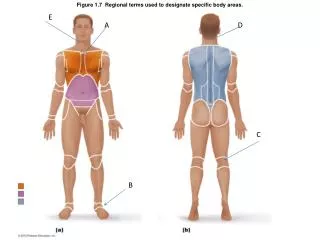
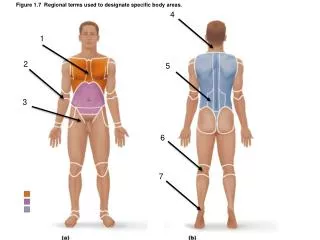
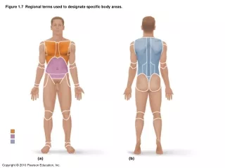
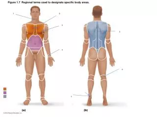
Figure 1.7 Regional terms used to designate specific body areas. 1. 2. 3. 5. 4. 6. 7. Figure 1.7a Regional terms used to designate specific body areas. thorax. abdomen. Figure 1.7b Regional terms used to designate specific body areas. dorsal.

eamon
Télécharger la présentation

Figure 1.7 Regional terms used to designate specific body areas.

An Image/Link below is provided (as is) to download presentation Download Policy: Content on the Website is provided to you AS IS for your information and personal use and may not be sold / licensed / shared on other websites without getting consent from its author. Content is provided to you AS IS for your information and personal use only. Download presentation by click this link. While downloading, if for some reason you are not able to download a presentation, the publisher may have deleted the file from their server. During download, if you can't get a presentation, the file might be deleted by the publisher.

E N D

Presentation Transcript


  1. Figure 1.7 Regional terms used to designate specific body areas. 1 2 3 5 4 6 7

  2. Figure 1.7a Regional terms used to designate specific body areas. thorax abdomen

  3. Figure 1.7b Regional terms used to designate specific body areas. dorsal

  4. Figure 1.8 Planes of the body with corresponding magnetic resonance imaging (MRI) scans. 8 9 10

  5. Figure 1.9 Dorsal and ventral body cavities and their subdivisions. 11 16 12 15 13 17 14

  6. Figure 1.9a Dorsal and ventral body cavities and their subdivisions. 18

  7. Figure 1.9b Dorsal and ventral body cavities and their subdivisions. 19 20

  8. Figure 4.3a Epithelial tissues. 21

  9. Figure 4.3b Epithelial tissues.

  10. Figure 4.3c Epithelial tissues.

  11. Figure 4.3d Epithelial tissues. 22

  12. Figure 4.3e Epithelial tissues.

  13. Figure 4.3f Epithelial tissues.

  14. Figure 4.8a Connective tissues.

  15. Figure 4.8b Connective tissues.

  16. Figure 4.8c Connective tissues.

  17. Figure 4.8d Connective tissues.

  18. Figure 4.8e Connective tissues. 23

  19. Figure 4.8f Connective tissues.

  20. Figure 4.8g Connective tissues.

  21. Figure 4.8h Connective tissues.

  22. Figure 4.8i Connective tissues.

  23. Figure 4.8j Connective tissues. 24

  24. Figure 4.8k Connective tissues.

  25. Figure 4.9a Muscle tissues.

  26. Figure 4.9b Muscle tissues.

  27. Figure 4.9c Muscle tissues.

  28. Figure 4.10 Nervous tissue. 25

  29. Figure 7.4a Anatomy of the anterior and posterior aspects of the skull. 26 27

  30. Figure 7.4b Anatomy of the anterior and posterior aspects of the skull. 28

  31. Figure 7.5a Bones of the lateral aspect of the skull, external and internal views. 29 30 31

  32. Figure 7.5c Bones of the lateral aspect of the skull, external and internal views. 32 33 34

  33. Figure 7.6a Inferior aspect of the skull, mandible removed. 35 36 37

  34. Figure 7.7a The base of the cranial cavity. 39 38 40

  35. Figure 7.13a Bones of the nasal cavity. 43 41 42

  36. Figure 7.13b Bones of the nasal cavity.

  37. Figure 7.14a Paranasal sinuses.

  38. Figure 7.14b Paranasal sinuses.

  39. Figure 7.15 The hyoid bone, anterior view.

  40. Figure 7.16 The vertebral column.

  41. Figure 7.19 Typical vertebral structures.

  42. Figure 7.20a The first and second cervical vertebrae.

  43. Figure 7.20b The first and second cervical vertebrae.

  44. Figure 7.20c The first and second cervical vertebrae.

  45. Figure 7.21a Posterolateral views of articulated vertebrae.

  46. Figure 7.21b Posterolateral views of articulated vertebrae. 44

  47. Figure 7.21c Posterolateral views of articulated vertebrae.

  48. Figure 7.22a The sacrum and coccyx.

  49. Figure 7.22b The sacrum and coccyx. 45

  50. Figure 7.23a The thoracic cage. 46

More Related