Download
1 / 62
650 likes | 1.5k Vues
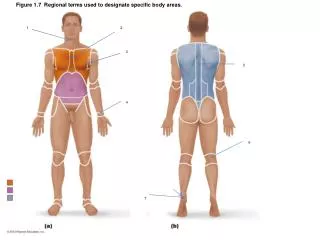
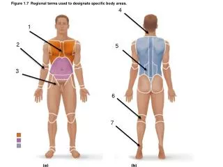
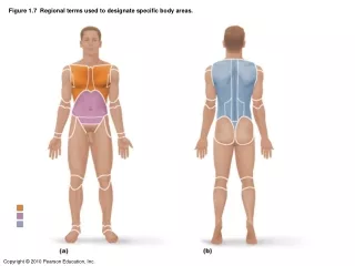
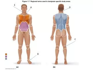
Figure 1.7 Regional terms used to designate specific body areas. E. A. D. C. B. A: Sternal B: Tarsal C: Popliteal D: Occipital E: Cervical. Figure 1.9b Dorsal and ventral body cavities and their subdivisions. A. B. A: Superior mediastinum B: Pleural Cavity.

E N D
Figure 1.7 Regional terms used to designate specific body areas. E A D C B
A: Sternal B: Tarsal C: Popliteal D: Occipital E: Cervical
Figure 1.9b Dorsal and ventral body cavities and their subdivisions. A B
A: Superior mediastinum B: Pleural Cavity
A: Visceral Pericardium B:Parietal pericardium C: Pericardial space with serous fluid
Figure 7.2b The skull: Cranial and facial divisions and fossae. B A
A: Middle Cranial Fossa B: Olfactory Foramina
Figure 7.26 The scapula. C B A
A: Glenoid Cavity B: SupraspinosusFossa C: Spine
Figure 7.27 The humerus of the right arm and detailed views of articulation at the elbow. A B C
A: Greater Tubercle B: Deltoid tuberosity C: Trochlea
A: Ilium B: Pubis C: Ischium
CHAPTER 4 • Slide 1: Simple squamous epithelium • Slide 2: Simple cuboidal epithelium • Slide 3: simple columnar epithelium • Slide 4: pseudostratified columnar epithelium • Slide 5: stratified squamous epithelium • Slide 6: transitional epithelium • Slide 7: Loose connective tissue areolar • Slide 8: Loose connective adipose • Slide 9: loose connective tissue reticular • Slide 10: dense regular connective tissue • Slide 11:dense irregular connective tissue • Slide 12: hyaline cartilage • Slide 13: elastic cartilage • Slide 14: fibrocartilage • Slide 15: skeletal muscle
Figure 10.21a Posterior muscles of the right hip and thigh. A B C D C
A: Gluteus Medius B: Adductor Magnus C: Semimembranosus D: Biceps Femoris E: Semitendinosus
Figure 10.20a Anterior and medial muscles promoting movements of the thigh and leg. C A D B E
A: Quadriceps Femoris B:Vastus Lateralis C: Pectineus D: Adductor longus E: VastusMedialis
A: Lateral ventrical B: 3rdVentrical C: 4Thventrical
Figure 12.22 Meninges: dura mater, arachnoid mater, and pia mater. A B C
A: Dura Mater B: Archanoid Mater C: Pia Mater
A: Hypothalamus B: Pineal Gland C: Medulla oblongata
Figure 15.3a Extrinsic eye muscles. B A C D E
A: Superior Rectus Muscle B: Superior Oblique C: conjunctiva D: Inferior Rectus Muscle E: Inferior Oblique
Figure 15.4a Internal structure of the eye (sagittal section). A B D C
A: Ciliary Body B:Choroid C: Retina D: Iris
A: spiral organ of corti B: Tectorial Membrane