Download
1 / 10
100 likes | 331 Vues
Crystal Structure of Rhodopsin : A G Protein-Coupled Receptor. Authors : Krzysztof Palczewski, Takasi Kumasaka, Tetsuya Hori, Craig A. Behnke, Hiroyuki Motoshima, Brian A. Fox, Isolde Le Trong,

E N D
Crystal Structure of Rhodopsin :A G Protein-Coupled Receptor Authors :Krzysztof Palczewski, Takasi Kumasaka, Tetsuya Hori, Craig A. Behnke, Hiroyuki Motoshima, Brian A. Fox, Isolde Le Trong, David C. Teller, Tetsuji Okada, Ronald E. Stenkamp, Masaki Yamamoto, Masashi Miyano Source : VOL 289 4 August 2000 Science Speaker : Ls02 871604 楊又穎
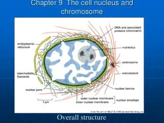
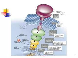
G protein : Heterotrimeric guanine nucleotide- binding protein associated with cytoplasmic face of the plasma membrane of mammalian cells , and involved in transmitting signals from receptors to intracellular pathways. Rhodopsin : a G protein-coupled receptor which is conjugated by opsin, phopholipid, retinal. Light sensitive, actived by light turn on the signal pathway that leads to vision.
Rhdopsin structure • Extracellular region • Transmembrane helices • Cytoplasmic surface
● Extracellular region -- mutation of Pro23 or Gln28 cause the eye disease retinities pigmentosa ● Transmembrane helices -- Helices H1,4,6,7 are bent at Pro residues -- The location of Gly90 is replaced by Asp causes night blindness (salt-bridge destabilization) -- Some hydrophobic binding sites
● Cytoplasmic surface -- C-IV loop, has a variety of synthesis peptides and their effect on the activation of G protein -- NPXXY motif -- hydrophobic enviroment ●11 cis-retinal chromophore -- retinylidene group -- trans form
Conclusion ● The crystal structure of rhodopsin reveals a highly organized heptahelical transmembrane bundle with 11-cis-retinal as a key cofactor involved in maintaining rhodopsin in the ground state. ● The structure gives information on the molecular mechanism of GPCR activation. ● A conformational change upon photoactivation of the chromophore that leads to rhodopsin activation and signal transduction.