Lecture 1: Crystallization Methods and Protein Crystal Properties
Lecture 1: Crystallization Methods and Protein Crystal Properties. Four major steps in crystallization. Obtain large amounts of pure protein samples Choose a protein buffer in which the protein is both soluble and stable

Lecture 1: Crystallization Methods and Protein Crystal Properties
E N D
Presentation Transcript
Lecture 1: Crystallization Methods and Protein Crystal Properties
Four major steps in crystallization • Obtain large amounts of pure protein samples • Choose a protein buffer in which the protein is both soluble and stable • Bring protein solution to supersaturation where spontaneous nucleation can take place • Crystal growth now begins
Solubility As a rule, protein solubility will usually increase as you add salt to your aqueous solution, then begin to decrease when the salt concentration gets high enough to compete with the protein for hydration (interaction with water molecules). HbCO (carboxyhemoglobin) solubility as a function of ionic strength in the presence of several different types of salts Diagram from the website of Alan Clark, Victoria University of Wellington, New Zealand http://www2.vuw.ac.nz/staff/alan_clark/teaching/index.htm
Supersaturation Supersaturation can be achieved by adding more of a substance (to a solution) than can normally be dissolved. This is a thermodynamically unstable state, achieved most often in protein crystallography by vapor diffusion or other slow evaporation techniques. Zone 1 - Metastable zone. The solution may not nucleate for a long time but this zone will sustain growth. It is frequently necessary to add a seed crystal. Zone 2 - Nucleation zone. Protein crystals nucleate and grow. Zone 3 - Precipitation zone. Proteins do not nucleate but precipitate out of solution. Diagram from the website for The University of Reading, Course FS460 Investigating Protein Structure and Function
Nucleation A phenomenon whereby a “nucleus”, such as a dust particle, a tiny seed crystal, or commonly in protein crystallography, a small protein aggregate, starts a crystallization process. Nucleation poses a large energy barrier, which is easier to overcome at a higher level of supersaturation. Common difficulties: 1. If supersaturation is too high, too many nuclei form, hence an overabundance of tiny crystals. 2. In supersaturated solutions that don’t experience spontaneous nucleation, crystal growth often only occurs in the presence of added nuclei or “seeds”.
Crystal Growth Adding single molecules to the surfaces of the nucleating lattice. Illustrated here through the work of Li and Nadarajah of The Macromolecular Crystallization Laboratory at the University of Toledo. AFM image of individual lysozyme molecules on the (110) face of a tetragonal crystal. (Li and Nadarajah) The growth steps and growth units of Lysozyme. The growth steps are at least bimolecular in height. The minimum growth unit for this step must be a tetramer corresponding to a single turn of the 43 helix as shown here.(Nadarajah) H. Li, M.A. Perozzo, J.H. Konnert, A, Nadarajah & M.L. Pusey, Acta Crystallographica, D55, 1023-1035 (1999).
Cessation of growth Caused by the development of growth defects or the approach of the solution to equilibrium. Mother liquor The solution in which the crystal exists - this is often not the same as the original crystallization screening solution, but is instead the solution that exists after some degree of vapor diffusion, equilibration through dialysis, or evaporation.
Major factors that affect crystallization 1) Purity of proteins 2) Protein concentration 3) Starting conditions (make-up of the protein solution) 4) Precipitating agent (precipitant) 5) Temperature 6) pH 7) Additives: Detergents, reducing agents, substrates, co-factors, etc.
Purity of proteins Sources of heterogeneity (other than unrelated proteins and nucleic acids as contaminants): • Partial proteolysis products • Oxidation of cysteines • Deamidation of Asn and Gln to Asp and Glu • Post-translational modifications • Oligomerization • Isoforms • Misfolded population • Structural flexibility
2) Protein concentration • Consistency andreproducibility are the major issues • with protein concentration - you should have a reliable • assay for determining the concentration. • Extinction coefficient for tryptophan • Bradford Assay (BSA is used as a standard) • E. Coli expression systems are crystallographers’ most commonly used method of obtaining protein. Problems can arise from low expression yields: • Cytotoxic - your protein is killing your E. coli • Unstable plasmid or mRNA • Protein is misfolded (coexpress with GroEL?) • Some common eukaryotic codons are rare in E. coli
3) Starting conditions (make-up of the protein solution) The main point is to KNOW what your starting conditions are for purposes of reproducibility.
4) Precipitating agent (precipitant) Salts Ammonium sulfate Sodium chloride Potassium phosphate Organic reagents MPD Isopropanol Polyethylene glycol PEG 4000 PEG 6000 PEG 8000
5) Temperature • Temperature affects protein stability and also the dynamics of how protein solution reaching supersaturated states. • Ideally: • An individual crystal screen should be kept at constant temperature • Each set of conditions should be screened at several temperatures • The easiest are 4 C and room temperature, also try 12 or 15 C
6) pH Surface charges affect “crystal packing”. (Crystal packing refers to the spatial arrangement of molecules within the crystal, particularly in reference to their relationships to one another.) Hydrophobic interactions are less important than electrostatic interactions in crystal packing.
7) Additives: • Sometimes you can increase the stability of your protein, • and/or the homogeneity of its conformation by having relevant additives present in the crystal screen: • Detergents • Reducing agents • Substrates • Co-factors • etc.
Still no crystals after thorough screening. Now what? • New constructs Deletion mutants Complexes with substrates Protein complex with Fab fragments Homologous proteins Fab
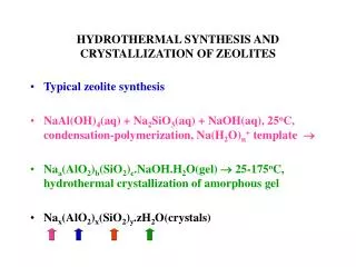
Crystallization of membrane proteins • The lipidic cubic phase method (Landau and Rosenbusch) • Cocrystallization with Fab fragments
Common Methods for Crystallization: Vapor Diffusion Slow Evaporation Dialysis
Hanging Drop Vapor Diffusion Most popular method among protein crystallographers. 1. Crystal screen buffer is the well solution (0.5 - 1 mL) 2. Drop (on siliconized glass cover slip) is 1/2 protein solution, 1/2 crystal screen buffer (6-10 L). So, the concentration of precipitant in the drop is 1/2 the concentration in the well. 3. Cover slip is inverted over the top of the well and sealed with vacuum grease (airtight). 4. The precipitant concentration in the drop will equilibrate with the precipitant concentration in the well via vapor diffusion.
Sitting Drop Vapor Diffusion Same basic principles as in hanging drop method, except the drop containing your sample sits on a bridge within the well. This allows for a larger sample size (20 - 40 L), however protein is frequently precious to the crystallographer, so there isn’t that much demand for a larger sample size.
Oil Immersion Micro Batch This method is rising rapidly in popularity- typical sample size 1-6 L Figure 1- Paraffin oil allows for little to no diffusion of water through the oil. This is a true batch experiment because all the reagents are present at a specific and relatively unchanging concentration. Figure 2- Al’s oil is a 1:1 mixture of silicon oil and paraffin oil which allows for evaporation through slow diffusion through the oil. This is an evaporation Method, and the concentration of the protein and reagents in the drop does increase over time.
Microdialysis Dialysis buttons can be purchased for a wide range ofsample sizes (~ 5 - 350 L). In the dialysis experiment, the sample is often introduced to high salt concentrations within the button that are allowed to equilibrate with lower salt concentrations in the buffer over time. This is known as a “salting-in” method. It exploits the fact that not only does protein solubility tend to decrease with very high ionic strengths, it also has a minimum at very low ionic strength.
View the Hampton Crystal Gallery to see in which labs each of these crystals originated and which biological molecule(s) they represent. http://www.hamptonresearch.com/stuff/gallery.html
The oscillation equipment Rotates the crystal about an axis () perpendicular to the x-ray beam (and normal to the goniometer). The diffraction pattern from a crystal is a 3-D pattern, and the crystal must be rotated in order to observe all the diffraction spots. This nice diagram also comes from Bernhard Rupp’s Crystallography 101 website: http://www-structure.llnl.gov/Xray/101index.html
Crystal Mounting Capillary tubes (Glass or Quartz) Cryo-loops (thin nylon)
Properties of protein crystals • Soft, easy to crush • Contain large solvent channels • Relatively large organic and inorganic molecules can diffuse inside • Anisotropic physical properties • Birefrigence due to anisotropic refraction indices • Ability to diffract X-ray due to regular spaced lattices