Advanced NanoPro Technology for Protein Signaling Molecule Profiling
40 likes | 166 Vues
The NanoPro Technology platform enables the detailed profiling of signaling molecules in precious small samples, such as stem cells and patient specimens. This innovative approach employs capillary isoelectric focusing combined with immunoassays, allowing for the separation, detection, and quantification of protein isoforms critical in cell signaling events. The methodology consists of loading samples, applying a separation voltage, immobilizing proteins, followed by targeted immunodetection using chemiluminescent imaging. This system reveals distinct phosphorylation patterns, offering insights into cancer signaling pathways and improving data reproducibility in research.

Advanced NanoPro Technology for Protein Signaling Molecule Profiling
E N D
Presentation Transcript
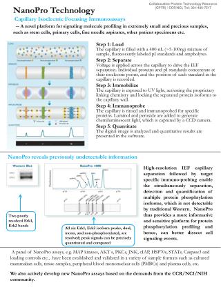
Collaborative Protein Technology Resource (CPTR) / CCR/NCI, Tel: 301-496-7517 NanoPro Technology Capillary Isoelectric Focusing Immunoassays -- A novel platform for signaling molecule profiling in extremely small and precious samples, such as stem cells, primary cells, fine needle aspirates, other patient specimens etc. • Step 1: LoadThe capillary is filled with a 400-nL (~5-100ng) mixture of sample, fluorescently labeled pI standards and ampholytes. • Step 2: SeparateVoltage is applied across the capillary to drive the IEF separation. Individual proteins and pI standards concentrate at their isoelectric points, and the position of each standard in the capillary is recorded. • Step 3: ImmobilizeThe capillary is exposed to UV light, activating the proprietary linking chemistry and locking the separated protein isoforms to the capillary wall. • Step 4: ImmunoprobeThe capillary is rinsed and immunoprobed for specific proteins. Luminol and peroxide are added to generate chemiluminescent light, which is captured by a CCD camera. • Step 5: QuantitateThe digital image is analyzed and quantitative results are presented in the software. NanoPro reveals previously undetectable information High-resolution IEF capillary separation followed by target specific immuno-probing enable the simultaneously separation, detection and quantification of multiple protein phosphrylation isoforms, which is not detectable by traditional Western. NanoPro thus provides a more informative and sensitive platform for protein phosphorylation profiling and hence, can better dissect cell signaling events. Two poorly resolved Erk1, Erk2 bands All six Erk1, Erk2 isoform peaks, dual, mono, and non-phosphorylated, are resolved; peak signals can be precisely quantitated and compared A panel of NanoPro assays, e.g. MAP kinases, AKT s, PKCs, JNK, cIAP, HSP70s, STATs, Caspase3 and loading controls etc., have been established and validated in a variety of sample formats such as cultured mammalian cells, tissue samples, peripheral blood mononuclear cells (PMBCs) and plasma cells, etc. We also actively develop new NanoPro assays based on the demands from the CCR/NCI/NIH community.
Collaborative Protein Technology Resource (CPTR) / CCR/NCI, Tel: 301-496-7517 NanoPro Application Examples Example 1:Divergent PKC delta phosphorylation pattern in LNCaP Cells revealed by NanoPro technology upon PMA and Bryostatin 1 stimulation. (Collaborator: Dr. Peter Blumberg) NanoPro 1000 Western Blot Erlotinib H2O Untreated EGFR pY1068 EGFR pY845 phospho-Erk1/2 • Upon PMA/ Bryostatin 1 treatment, several peaks shifted to the more acidic (Lower pI) regions suggesting multiple-site phosphorylation. The event is able to be detected by a single pan-anti-PKCdelta antibody; • A more simple peak pattern was observed in Bryostatin 1 treatment, which may contribute to the different biological responses observed for Bryostatin 1 and PMA. • Phospho-specific antibody is needed to detect PKC activation; • Cannot distinguish differences between complex phosphorylation patterns of PKC upon PMA and Bryostatin 1 treatment Actin Untreated Example 2:NanoPro technology enables the detection and distinction of cancer cell specific signals from stroma cells in xenograph tissue samples. (Collaborator: Dr. Giuseppe Giaccone ) NanoPro 1000 Western Blot H2O Erlotinib Mouse Erk1 Human Erk1 Cannot distinguish human and mouse isoforms, thus not able to differentiate signals from human cancer cells and interference from mouse stroma cells Separates human Erk1 from mouse Erk1 by pI difference, demonstrating that Erlotinib inhibition on Erk phosphorylation is specific to human cancer cells
Collaborative Protein Technology Resource (CPTR) / CCR/NCI, Tel: 301-496-7517 Simple Western System – Simon Capillary based automated Western system – all steps computer programmed, including sample loading, size-based protein separation, immunoprobing, washing, detection and data analysis; highly quantitative data with excellent reproducibility. Work Flow Assay Reproducibility Proteins are loaded into the capillary automatically and separated by size as they migrate through a stacking and separation matrix. The separated proteins are then immobilized to the capillary wall via a proprietary, photo-activated capture chemistry. Target proteins are identified using a primary antibody and immunoprobed using an HRP-conjugated secondary antibody and chemiluminescent substrate. The resulting chemiluminescent signal is detected and quantified. Simon simple Western assays are fully automated, which minimizes the variability caused by manual processes in traditional Western, such as inconsistencies in protein transfer etc. Examples shown above demonstrate excellent intra-assay, inter-run, and inter- person data reproducibility in Simon system.
Collaborative Protein Technology Resource (CPTR) / CCR/NCI, Tel: 301-496-7517 Simon Application Example -- Simon also provides a platform for precise quantification and direct comparison of protein isoforms in cell lysate, and thus a method for correlating protein quantity with functions Endogenous Erk2 Measurement in LNCaP Lysate Serial dilutions of GST-Erk2 were spiked into cell lysate, and detected simultaneously with endogenous Erk2 GST-Erk2 sandard curve GST-ERk2 Linear regression analysis for endogenous Erk2 calculation ERk2 8.26pg Erk2 in 16.5 ng LNCaP Lysate 0.050% of total protein • NanoPro Technology Summary • • Conserve precious samples — characterize proteins directly in limited cellular sub-populations • • Detect multiple phosphorylation states — measure subtle shifts in phospho-isoform distributions • • Higher data quality — precise, automated assays and quantitative digital data analysis provide more accurate and reproducible results • Simon Technology Summary • Fully automated, gel free and blot free • Highly quantitative data and excellent reproducibility Contact Information Jinqiu Chen, PhD (Program Manager) chenj13@mail.nih.gov, 301-496-7517; Michelle Herrmann, MS herrmannma@mail.nih.gov, 301-594-3749