CIRCULAR DICHROISM SPECTROSCOPY
CIRCULAR DICHROISM SPECTROSCOPY. INTRODUCTION. Types of Polarized Light. Light is an electromagnetic radiation where the electric vector (E) and the magnetic vector (M ) are perpendicular to each other .

CIRCULAR DICHROISM SPECTROSCOPY
E N D
Presentation Transcript
Types of Polarized Light Light is an electromagnetic radiation where the electric vector (E) and the magnetic vector (M) are perpendicular to each other. Linearly or plane-polarized lightis obtained by passing light through a polarizer that transmits light with only a single plane of polarization(components of the E vector that are parallel to the axis of the polarizer). If the E vectors of two electromagnetic waves are ¼ wavelength out of phase and perpendicular to each other, the vector that is the sum of the E vectors of the two components rotates around the direction of propagation so that its tip follows a helical path. Such a light is called circularly polarized light.
Types of Polarized Light Circularly polarized light can be left-handed or right-handed. Right-handed circularly polarized light rotates clockwise.
Optical Isomers Two non-super imposable mirror images of a molecule are known as Optical Isomers or Enantiomers. Super imposable Mirror image Non-super imposable Mirror image
Optical Rotation It is the turning of the plane of linearly polarized light about the direction of motion as the light travels through certain materials.
Optical Rotary Dispersion It is the variation in the optical rotation of a substance with a change in the wavelength of light. Optical Rotary Dispersion Spectroscopy is a technique for measuring the ability of light, passing through an optically active substance, to rotate the plane of polarization, as a function of wavelength. However, such substances may also absorb the plane-polarized radiation at certain wavelengths. Here, the chromophore (group which is responsible for the colour of a compound) is termed as an optically active chromophore or chiral centre, as it can be a part of only a complex molecule. The technique of ORD has been largely replaced by Circular Dichroism (CD) Spectroscopy as it gives better information about the 3-D structure of macro-molecules containing chiral centres.
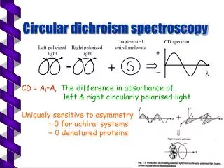
Circular Dichroism In addition to changing the plane of polarization, an optically active sample also shows unusual absorption behaviour. Left- and right-handed polarized components of the incident light are absorbed differently by the sample, which yields a difference in the absorption coefficients. This latter difference is called Circular Dichroism (CD). “Dichroism” is used to denote direction-dependent light absorption.
Circular Dichroism Definition : Circular Dichroism (CD) Spectroscopy measures the differences in the absorption of left-handed polarized light versus right-handed polarized light.
Circular Dichroism For plane-polarized light : Chiral (centre) molecules interact differently with polarized light due to the asymmetry in its structure. The R and L waves are differentially absorbed and refracted and result in a beam in a different plane.
Circular Dichroism For circularly-polarized light : When this light passes through the sample, the resultant beam is a recombination of the R and L components, giving an emergent beam of elliptically polarized light.
Circular Dichroism In CD spectroscopy, this ellipticity of the light is measured. It is given by – θ = 2.303ΔA = 33 ΔA (degrees) Here, ΔA is the difference in absorption between R and L components.
Circular Dichroism CD Spectrum : It is a plot of ellipticity versus wavelength.
Circular Dichroism The basic layout of a CD spectrometer follows that of a single-beam UV absorption spectrometer. Generally, left and right circularly polarized light passes through the sample in an alternating fashion. This is achieved by an electro-optic modulator which is a crystal that transmits either the left- or right-handed polarized component of linearly polarized light, depending on the polarity of the electric field that is applied by alternating currents. The photomultiplier detector produces a voltage proportional to the ellipticity of the resultant beam emerging from the sample.
Circular Dichroism Practical Considerations : A clean quartz cuvette Buffers with low concentrations of additives Filtered solutions should be used to avoid any turbidity of the sample To calculate specific ellipticities and to compare the CD spectra of different samples with each other, the concentration of the sample must be known.
Circular Dichroism The major application of CD is the study of conformation of biological macromolecules. Proteins : The main application for protein CD spectroscopy is the verification of the adopted secondary structure. Information can be gained about the relative proportions of secondary structure, α-helical,β-sheet and random coils in solution.
Circular Dichroism Nucleic Acids : For calculating the CD spectrum of a single strand of DNA from the nearest known neighbour frequency. To study structural changes in nucleic acids (e.g. Loss of helicity of single-stranded DNA as a function of temperature and pH)