Science 30 Circulation and Immunity
Science 30 Circulation and Immunity. Introduction: Launch Lab – Watching Blood Flow. Procedure: Watch the video clip showing blood circulation in the tail of a gold fish. Answer the analysis questions. Circulation in a Goldfish Tail. Structures of the Circulatory System.

Science 30 Circulation and Immunity
E N D
Presentation Transcript
Introduction: Launch Lab – Watching Blood Flow • Procedure: • Watch the video clip showing blood circulation in the tail of a gold fish. • Answer the analysis questions. Circulation in a Goldfish Tail
Structures of the Circulatory System • The circulatory system is the transportation system of the body. • 3 Main Functions • Transports gases • Regulates internal temperature • Protect against blood loss and infection
Major Components • 3 Major Components: • The heart – an organ that pushes blood through the body with its pumping action. • The blood vessels – serve as the “roadways” through which the blood moves. • The blood – carries nutrients, oxygen, carbon dioxide, water, wastes and many other materials throughout the body.
The Cardiovascular System • Together the heart and the blood vessels are known as the cardiovascular system.
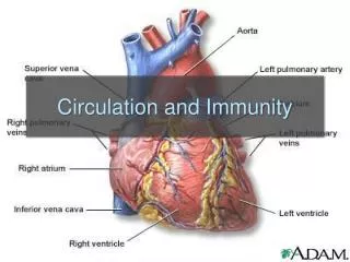
The Structure of the Heart • Located on the left side of the chest • The heart: • Pumps blood • Separates oxygen rich and poor blood • Keeps blood flow in one direction
The walls of the heart are made up of cardiac muscle tissue • The contractions of the cardiac muscle tissues are rhythmical and involuntary
The human heart has 4 chambers: • Left and right atria – fill with blood from body or lungs • Left and right ventricles – pump blood to the body or lungs • The left and right side is separated by a thick wall called the septum
Blood Flow through the Heart • Oxygen-poor blood enters the heart from the vena cava • Superior vena cava collects blood from upper body • Inferior vena cava collects blood from below the chest
vena cava R atrium R ventricle pulmonary arteries lungs L atrium L ventricle aorta body
Valves of the Heart • 4 valves control direction of flow: • Right atrioventricular valve: • Separates right atrium and ventricle • Tricuspid; made of 3 flaps • Left atrioventricular valve: • Separatesleft atrium and ventricle • Bicuspid; made of 2 flaps
Chordae tendinae: • hold atrioventricular valves in place • prevent valves from being pushed back into atria • Semilunar valves control the flow of blood into the aorta and pulmonary artery
Using blue and red pencils or pens, diagram the pathway of blood through the heart using arrows. Use blue to represent deoxygenated blood and red for oxygenated blood.
Blood enters the right atrium through the ________________. • Blood flows from the right atrium into the right ventricle through the _________________. • Blood is pumped from the right ventricle into the pulmonary trunk that splits into the right and left _____________________. • Blood returns from the lungs by way of the right and left ____________________. • Blood enters the ____________________ when it returns from the lungs. • Blood flows past the ________________________ as it enters the left ventricle. • The left ventricle pumps out past the ______________________ into the aorta.
The Structure of Blood Vessels • Three types of blood vessels: • Arteries: • carry oxygen-rich blood away from the heart • thick and highly elastic walls • Veins: • carry oxygen-poor blood towards the heart • thinner walls and large inner circumference • have one-way valves to prevent back flow
Capillaries: • network of fine vessels where gas and nutrient transfer to tissues occurs. Capillaries join arteries to veins • Capillary walls are a single cell thick
Label the diagram • Use blue and red to indicate whether the vessel carries oxygenated blood (red), deoxygenated blood (blue) or both.
Compare the structure and function of the three types of blood vessels.
The Beating Heart The stimulus that triggers a heartbeat is an electrical signal that originates from within the heart. The sinoatrial (SA) node is a bundle of specialized muscle tissue that stimulates the muscle cells to contract and relax rhythmically Located in the wall of the right atrium SA node is also called the “pacemaker” because it sets the pace for cardiac activity
The SA node generates an electrical signal that spreads over the two atria simultaneously. • As the atria contract, the signal reaches the atrioventricular (AV) node. • The AV node transmits the electrical signal through the bundle of specialized fibres called the bundle of His. • The bundle of His relays the signal through two bundle branches that divide into fast-conducting Purkinje fibres • Purkinje fibres initiate the simultaneous contraction of all cells of the right and left ventricles Heartbeat signal
ECG • Changes in voltage of the heart can be measured using an electrocardiogram (ECG) • An ECG measures the electrical activity of the heart as it contracts and relaxes • Irregular spikes or changes in the spacing of waves can be used to diagnose different heart conditions
Blood Pressure • Blood passing through vessels exerts pressure against vessel walls, called blood pressure • Changes in blood pressure corresponds to phases of the heartbeat • Maximum pressure is called systolic pressure, during ventricle contraction • Lowest pressure is called diastolic pressure, occurs just before the ventricles contract • Sphygmomanometer (blood pressure cuff) measures blood pressure
Cardiac Output and Stroke Volume • Cardiac output is the amount of blood pumped by the heart, measured in mL/min • Cardiac output is an indicator of the level of oxygen delivered to the body and the amount of work the body’s muscles can perform • Heart rate is the number of heartbeats per minute • Stroke volume is the amount of blood forced out of the heart with each heartbeat. • cardiac output = heart rate x stroke volume
Stroke volume depends on: • How easily the heart fills with blood, related to “stretchiness” of ventricular walls and volume returning to heart from veins • How readily the heart empties, related to the strengthof the ventricular contraction • Average person has a stroke volume of 7o mL and a resting heart rate of 70 beats/min • Cardiac output = 70 mL x 70 beats/min = 4900 mL/min • Average person has 5 L of blood in their body; the total volume of blood circulates through the heart about once every minute.
Pathways of the Circulatory System • The circulatory system has 3 pathways: • Pulmonary pathway – transports blood between the heart and the lungs • Systemic pathway – moves blood from the left ventricle to the tissues and back to the right atrium • Coronary pathway– provides blood to the heart itself
Tracing the Pathways… • Tracing the pathway of blood beginning in right atrium… pulmonarypulmonary systemic systemic (to lungs) (to heart) (to tissues) (to heart)
Tracing the pathway of blood through coronary… • Heart does not use blood inside chambers to get nutrients/ remove wastes; walls are too thick fordiffusion • The heart is covered in a network of vessels • Oxygenated blood is supplied from the coronary artery coming off of the aorta • Deoxygenated blood enters the right ventriclebefore heading to the lungs.
Cardiovascular Disorders and Treatments • Cardiovascular disease is the leading cause of death for Canadians • Can be reduced with lifestyle changes: • not smoking • Eating healthy diet • Exercising • Atherosclerosis: • thickening of artery walls • Elasticproperties diminished
Atherosclerosis: • Build-up of fatty deposits on wall of artery • Blood flow is decreased, blood pressure is increased • Especially dangerous in arteries of heart, neck, brain, legs, kidneys • Treated withaspirinto prevent platelets from sticking together (clots) • Treated with medication tobreakdown existing clots Artherosclerosis
Surgical treatments such as angioplasty; permanent stent is inserted into artery to “re-open” passage way Angioplasty
Coronary bypass operation – healthy vessels are taken from elsewhere in the body and used to create a new pathway around a blocked vessel. Coronary Bypass Surgery
Congenital Heart Defects • Some heart defects are congenital, they are present since birth • Common congenital defects: • wallsdividing chambers • valvesof the heart • structure of the blood vesselsnear the heart
Heart murmur – describes any misflow of blood through the heart • Ie. One or more of the valves not opening properly • Valve defects can beheard with a stethoscope as a whooshing or rasping sound (caused by blood “leaking” through the valve • Can be also be diagnosed with CT scan, CAT scan, MRI scan by creating a 3D image of the organ Bicuspid/ Mitral valve Regurgitation Heart Transplant
Blood • Average 70kg person has 5L of blood. • 55% of blood is fluid,45% is blood cells. • Plasma = fluid; 90% water and 10% proteins, vitamins, glucose... • There are 3 groups of proteins in plasma: • Albumins: draws water back into capillaries • Globulins: produces antibodies • Fibrinogens: used to clot blood.
A. Red Blood cells (erythrocytes) • Make up 99% of blood cells. • Main function = transport oxygen (each rbc can have 4 oxygen molecules). • Respiratory pigment hemoglobin allows rbc’s to carry oxygen ( 70x more than without hemoglobin)- gives red color! • Without hemoglobin, life can be maintained for 4.5s; with = 5 minutes.
biconcave shape to maximize SA for gas exchange. • Enucleated = no nucleus when mature, allows for more oxygen to be carried. Bone marrow produces rbc’s, can not reproduce without a nucleus.
Bone marrow • Erythropoiesis= rbc production ( occurs in bone marrow). • Rbc’s begin as stem cells, divide and shrink- losing nucleus. • Age of rbc’s is monitored by wbc’s; when rbc is 120 days, it releases hemoglobin and is transported from the cell. • Iron (globin) is recycled and used in new rbc’s; heme is converted into bile pigments (bilirubin).
B. White blood cells (Leukocytes) • Around 1% of blood. • Responsible for: housekeeping/ defence. • Have a nucleus. • Made in bone marrow; last 13- 20 days. • Destroy invaders by: • Phagocytosis: move towards microbe. Enzymes released digest leukocyte and microbe; fragments = pus.