Download

1 / 10
110 likes | 288 Vues
HIV GP120 Envelope Protein. Objectives : Observing consensus sequences of different strains Structure analysis . GP120 Function. GP120 involved in infection of human T-cells. HIV TYPES. HIV Type I. SIV. HIV Type II. HIV Type III. Human viruses. Simian virus. GP120 Sequence analysis.

E N D
HIV GP120 Envelope Protein • Objectives : • Observing consensus sequences of different strains • Structure analysis
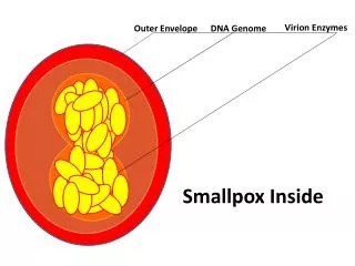
GP120 Function GP120 involved in infection of human T-cells
HIV TYPES HIV Type I SIV HIV Type II HIV Type III Human viruses Simian virus
GP120 Sequence analysis Sequence analysis shows a high mutation rate with preserved large consensus regions not only between strains, but also between different types
GP120 Sequence analysis HIV Type I shows additional sequence of 7 aa in it’s consensus sequence in a consensus region in acids 27-33 consensusLWVTVFYGVPVWKEATTTLFCATDNK-------NTWATHACVPTDPDYQEVELNNVTENF 53 HIV1LWVTVYYGVPVWHDADPVLFCASDAKahsteahNIWATQACVPTDPSPQEVFLPNVIESF 92
Two structures of GP120-CD4 from HIV 1 were analyzed Hiv-1 Yu2 isolate Hiv-1 Hxbc2 isolate
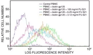
Difference between the proteins The similarity is shown, using protein blast Score = 566 bits (1460), Expect = e-161Identities = 276/321 (85%), Positives = 289/321 (89%), Gaps = 8/321 (2%)
Structure of conserved region GP120 Hxbc2 isolate Preserved region marked in yellow CD4
Docking of GP120 Clustered solution of Dimers With current approach GP120 exists as a trimers or tetramers on the membrane surface. According to J. Biol. Chem., Vol. 275, Issue 45, 35137-35145, November 10, 2000
Conclusions • Preserved sequences found • GP120-CD4 theoretical docking performed, clustered solutions found • The results provide a basis for further research