
Oxygen Debt vs. Recovery Oxygen
E N D
Presentation Transcript
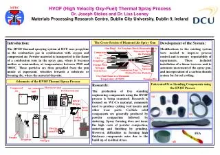
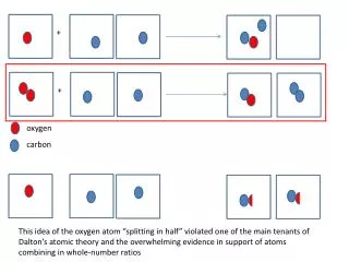
1. Oxygen Debt vs. Recovery Oxygen Oxygen Debt (AV Hill) stated most lactic acid formed during exercise was converted to glycogen (~80%),
Margaria introduced concept of �Alactacid and lactacid debt components
Contemporary termed �Recovery Oxygen� or Excess post-exercise oxygen consumption� (EPOC)
Fast component and slow component
2. Fast Component Phosphagens (ATP & PC)
Elevated oxygen consumption during this time period fuels the body�s need for:
restoring myoglobin w/oxygen
restoring blood levels of O2
O2 cost of breathing
Increased heart rate (myocardial O2
Replenish phosphagens
Magnitude of Fast Component volume is related to previous exercise intensity Include VO2 curve for steady state and for recovery.Include VO2 curve for steady state and for recovery.
3. Fast Component Recovery Rapid recovery of phosphagens at start of recovery & later slows down
70% - 30 seconds
100% - 3-5 minutes
PC recovery
84% restored in 2 minutes
89% restored in 4 minutes
4. Replenishment of Energy Stores Restoration of ATP + PC
Half-time = 30 sec; 1 min = 3/4�s; 1.5 min = 7/8; 3 min = 63/64
Muscle glycogen
.7 g/kg body wt/hr, carbohydrates with high glycemic index -
On average 5% of muscle glycogen used during exercise is resynthesized each hour after exercise; minimal 20 hrs to restore muscle
Lactic acid removal Provided 600 grams of CHO are consumed.
During successive days of competition or intense training, athletes hould consume approximately 100 g of carbohydrate within 15-30 min after exercise followed by an additional 100 g feedings every two to four hours.
60% replenished after 10 hours providing a high carbohydrate intake
Liquid, solid, simple sugar, complex carbohydrate feedings appear to be equally effective.
Provided 600 grams of CHO are consumed.
During successive days of competition or intense training, athletes hould consume approximately 100 g of carbohydrate within 15-30 min after exercise followed by an additional 100 g feedings every two to four hours.
60% replenished after 10 hours providing a high carbohydrate intake
Liquid, solid, simple sugar, complex carbohydrate feedings appear to be equally effective.
5. O2 - Myoglobin Stores Found in skeletal muscle
Store for oxygen
Involved functionally in transfer of oxgyen
Inside the cell to inside the mito
11.2 ml/kg of muscle or ~500 ml
Functional significance related to intermittent exercise - rapidly restored following short bout of exercise
6. Slow Component of Recovery Elevated body temperature - ? metabolic activity
Oxygen cost of ventilation
Oxygen cost of ? myocardial activity
? Na-K pump
Glycogen resynthesis
Oxidation of lactic acid Slow component is relatively unaffected by a change in exercise intensity or duration until a threshold of a combined intensity (80%) and duration (20 min) is presented
Hagberg (1980) stated that 60-70% of slow component oxygen can be accounted by effect of temperature.Slow component is relatively unaffected by a change in exercise intensity or duration until a threshold of a combined intensity (80%) and duration (20 min) is presented
Hagberg (1980) stated that 60-70% of slow component oxygen can be accounted by effect of temperature.
7. Exercise � Related factors that cause extra O2 consumption Temperature � effects mitochondrial O2 consumption, leading to decline in energy �trapping efficiency� (Brooks, 2000)
BMR? 13% per degree Celsius increase in body temp
Sympathetic stimulation � increases metabolic rate of numerous tissues
8. Lactic Acid Removal Rest recovery - half-time = 25 min.; 75 min for near complete recovery (95%)
Exercise recovery - 30-45% of VO2max untrained; 50-65% VO2max trained; Stainsby, 1970.
Half-time = 10 � 11 minutes
Complete recovery = ~ 25 minutes (Bonen & Belcastro, 1976)
9. EPOC � Slow Recovery Rest recovery - half-time = 25 min.; 75 min for complete recovery
Exercise recovery - 30-45% of VO2max untrained; 50-65% VO2max trained; - Stainsby, 1970.
10. Muscle Glycogen Resynthesis Full repletion of muscle glycogen requires several days and depends on:
Type of exercise causing depletion
Amount of dietary carbohydrate consumed during recovery period Muscle glycogen depletion can be caused by: continuous endurance-like activities & intermittent, exhaustive activities)
The studies from your text tended to be of 1-hr duration.
Muscle glycogen depletion can be caused by: continuous endurance-like activities & intermittent, exhaustive activities)
The studies from your text tended to be of 1-hr duration.
11. Continuous Endurance Exercise Insignificant amount of muscle glycogen resynthesized in 1st 1-2 hours of recovery
Complete restoration of muscle glycogen w/ high diet of CHO requires ~46 hours
W/out high CHO diet, small quantity of muscle glycogen resynthesized during 5 d
Rapid replenishment occurs during first several hours of recovery When food is available and physical activity is low, the hormonal situation (elevated insulin and low catecholamines) increases the activity of the glycogen storing enzyme glycogen synthetase
To speed the replenishment of carbohydrates after a hard period of training or competition, it is wise to begin consuming carbohydrate-rich foods ASAP after exercising.
Recommend consuming 100g CHO within the first 15-30 minutes of recovery.
Consume high to oderate glycemic carbohydrates every two hours until 500 g are consumedWhen food is available and physical activity is low, the hormonal situation (elevated insulin and low catecholamines) increases the activity of the glycogen storing enzyme glycogen synthetase
To speed the replenishment of carbohydrates after a hard period of training or competition, it is wise to begin consuming carbohydrate-rich foods ASAP after exercising.
Recommend consuming 100g CHO within the first 15-30 minutes of recovery.
Consume high to oderate glycemic carbohydrates every two hours until 500 g are consumed
12. Intermittent, Short Duration Exercise A significant amount of muscle glycogen resynthesized in 30 min - 2 hrs with no food
Complete resynthesis of muscle glycogen does not require a greater than normal intake of carbohydrate
Complete resynthesis of muscle glycogen required a 24-hour recovery period when a normal or high CHO diet is consumed
Muscle glycogen is most rapid during first several hours of recovery More rapid resynthesis takes place if the person remains inactive during the recovery period.
With optimal CHO intake, glycogen stores are replenished at a rate of 5-7% per hour.
Therefore, under the best conditions, it will take at least 20 hours to reestablish glycogen stores after a glycogen-depleting bout of exercise
Bangsbo has found similar findings regarding the partial restoration of muscle glycogen during 1st hour of recovery.More rapid resynthesis takes place if the person remains inactive during the recovery period.
With optimal CHO intake, glycogen stores are replenished at a rate of 5-7% per hour.
Therefore, under the best conditions, it will take at least 20 hours to reestablish glycogen stores after a glycogen-depleting bout of exercise
Bangsbo has found similar findings regarding the partial restoration of muscle glycogen during 1st hour of recovery.
13. High & Moderate Glycemic Foods High glycemic foods - contain glucose, sucrose, cane maple, corn syrup, bagel, white potato, honey, carrots
Moderate glycemic foods - whole grain bread, spaghetti, oatmeal, orange
Glycemic index of food is determined from the integrated rise in blood glucose following consumption of a standard amount (50-100g) compared to glucose
Jenkins et al., 1981 Glycemic effect � describes the effect of food on blood glucose; how quickly glucose is absorbed after a person eats, how high blood glucose rises and how quickly it returns to normal
Slow absorption, a modest rise in blood glucose, and a smooth return to normal are considered desirable;
Fast absorption, a surge in blood glucose, and an overreaction that forces glucose below normal are undesirable.
Glycemic index provides a measure of the rapidity with which sugar enters into blood following food consumption.Glycemic effect � describes the effect of food on blood glucose; how quickly glucose is absorbed after a person eats, how high blood glucose rises and how quickly it returns to normal
Slow absorption, a modest rise in blood glucose, and a smooth return to normal are considered desirable;
Fast absorption, a surge in blood glucose, and an overreaction that forces glucose below normal are undesirable.
Glycemic index provides a measure of the rapidity with which sugar enters into blood following food consumption.
14. Skeletal Muscle Functional Anatomy Intact skeletal muscle organ
Composed of muscle fascicles
Composed of muscle fibers The intact skeletal muscle is composed of muscle fascicles or bundles, which in turn are composed of muscle fibers.
Because each fiber is a single multinucleated cell, the term cell and fiber are used interchangeably.
Fibers in turn, are composed of myofibrils arranged in parallel.
Myofibrils are composed of sarcomeres arranged in series.
Sarcomeres are the basic contractile units of skeletal muscle - composed of interdigitating thick and thin myofilaments
The intact skeletal muscle is composed of muscle fascicles or bundles, which in turn are composed of muscle fibers.
Because each fiber is a single multinucleated cell, the term cell and fiber are used interchangeably.
Fibers in turn, are composed of myofibrils arranged in parallel.
Myofibrils are composed of sarcomeres arranged in series.
Sarcomeres are the basic contractile units of skeletal muscle - composed of interdigitating thick and thin myofilaments
15. Different Kinds of Motor Units Aerobic Type Fibers
Type I, red, slow-twitch (ST), or slow oxidative (SO)
Anaerobic Type Fibers
Type II, white, fast-twitch (FT), or fast-glycolytic (FG)
IIA - fast-oxidative-glycolytic (FOG)
IIB - fast-glycolytic (FG)
IIC - Unclassified Myogenic factors have been found in muscle fibers that vary and affect the properties specific to the fiber.
That is, fibers have different forms of myofibrillar and other proteins = isoforms
Thus, fibers have different speeds and power production during contraction.
One of the main differences is the myosin heavy chains.
Type II fibers have a troponin with a higher Ca++ binding capacity and a faster process of cross-bridge attachment.Myogenic factors have been found in muscle fibers that vary and affect the properties specific to the fiber.
That is, fibers have different forms of myofibrillar and other proteins = isoforms
Thus, fibers have different speeds and power production during contraction.
One of the main differences is the myosin heavy chains.
Type II fibers have a troponin with a higher Ca++ binding capacity and a faster process of cross-bridge attachment.
16. Basement Membrane Located adjacent to sarcolemma
Loose collection of glycoprotein and collagen - not lipid bilayer
Freely permeable to protein, solute, metabolites
Wedged between B.M. and sarcolemma are cells known as satellite cells
Important for growth & development
Adaptive capacity to training & disuse
Recovery from exercise induced damage Normally, satellite cells are dormant, but under conditions of stress or injury they are essential for the regenerative growth of new fibers.
Satellite cells can migrate from one location to another area of higher need within a muscle fiber.
Can undergo cell division to produce additional satellite cells.
These cells migrate accross the sarcolemma into cytoplasm/cytosol, where they recognize each other, align and fuse into a myotube.
Myotube is an immature muscle cell/fiber
The multinucleated myotube will then differentiate into a mature fiber.
In case of serious injury or neuromuscular disease, satellite cells are critical for regenerative growth
What about hypertrophy of existing cells?
Satellite cells are activated when muscle fibers hypertrophy.
It remains to be determined how much this occur when muscles undergo resistance training.Normally, satellite cells are dormant, but under conditions of stress or injury they are essential for the regenerative growth of new fibers.
Satellite cells can migrate from one location to another area of higher need within a muscle fiber.
Can undergo cell division to produce additional satellite cells.
These cells migrate accross the sarcolemma into cytoplasm/cytosol, where they recognize each other, align and fuse into a myotube.
Myotube is an immature muscle cell/fiber
The multinucleated myotube will then differentiate into a mature fiber.
In case of serious injury or neuromuscular disease, satellite cells are critical for regenerative growth
What about hypertrophy of existing cells?
Satellite cells are activated when muscle fibers hypertrophy.
It remains to be determined how much this occur when muscles undergo resistance training.
17. A Single Multinucleated Cell Muscle Cell = Muscle Fiber
Fibers composed of myofibrils
Myofibrils are composed of Sarcomeres
Sarcomeres are composed of thick & thin myofilaments
18. Connective Tissue Membranes Epimysium
Perimysium
Basement membrane, endomysium, basal lamina The basement membrane is not a membrane in the usual sense. Rather than having the normal structure of a lipid bi-layer, the basement membrane is a loose collection of glyco-proteins and collagen network. It is freely permeable to proteins, solutes and other metabolites.
An additional thin elastic membrane is found just beneath the basement membrane, and the terms plasma membrane or sarcolemma are used interchangeably.
The sarcolemma is much more selective to ions, solutes and substrates crossing it.
Critical for the following reasons:
1. Maintain acid - base balance in the fiber
2. Propagates the action potential/nerve impulse
3. Transports metabolites from the blood to the cytosol
4. At neuromuscular junction, a more elaborate region of functional folds
The basement membrane is not a membrane in the usual sense. Rather than having the normal structure of a lipid bi-layer, the basement membrane is a loose collection of glyco-proteins and collagen network. It is freely permeable to proteins, solutes and other metabolites.
An additional thin elastic membrane is found just beneath the basement membrane, and the terms plasma membrane or sarcolemma are used interchangeably.
The sarcolemma is much more selective to ions, solutes and substrates crossing it.
Critical for the following reasons:
1. Maintain acid - base balance in the fiber
2. Propagates the action potential/nerve impulse
3. Transports metabolites from the blood to the cytosol
4. At neuromuscular junction, a more elaborate region of functional folds
19. Sarcomeres Mid-Portion - A-Band
Outer ends - I-Bands
H - Zone - bisected by M line
Z - Disks or Z - Lines
A-band - area that appears dark, composed of myosin. isotropic
I-band - outer ends of each sarcomere appear light, primarily actin
H-zone - central portion of A-band, more lightly stained - no actin
associated with myosin.
M - line contains proteins that keep the sarcomere in proper spatial orientation as it lengthens and shortens
Z - lines - located at the end of each sarcomere
The I-band and H-zone are less dense than other areas as there is no overlap of thick and thin filaments; this allows for greater penetration of light when examined with a microscope.
20. Contractile & Regulatory Proteins 20% of skeletal muscle comprised of protein
12% myofibrillar protein
8% enzymes, membrane proteins, transport channels and other proteins
21. Myofibrillar Fraction of Skeletal Muscle Myosin
Actin
Tropomyosin
Troponin
C Protein
M-line protein
Alpha Actinin C protein - part of the thick filament, it is involved in holding the tails of myosin in a correct spatial agreement
M - Line protein functions to keep the thick and thin filament in their correct spatial arrangement
Alpha Actinin - attaches actin filaments together at the Z-disk.
C protein - part of the thick filament, it is involved in holding the tails of myosin in a correct spatial agreement
M - Line protein functions to keep the thick and thin filament in their correct spatial arrangement
Alpha Actinin - attaches actin filaments together at the Z-disk.
22. Myosin Thick filament
Light meromyosin (LMM)
Heavy meromyosin (HMM)
S-1 and S-2 Subfragments
Together form the crossbridge S-2 portion of HMM projects out at an angle from LMM =tail, and the
S-1 portion is the globular head that can bind to actin - binding site for ATP and storage of energy
S-2 portion of HMM projects out at an angle from LMM =tail, and the
S-1 portion is the globular head that can bind to actin - binding site for ATP and storage of energy
23. Myosin Myosin protein plays key role in development of muscular force & velocity of contraction
Myosin is a hexameric (six-component) molecule
1 pair Myosin heavy chains (MHCs) (high molecular weight)
2 pairs of light chains (LCs)
MHC protein molecules called isoforms These MHC isoforms have slight variations in their amino acid composition, particularly in the head regions (S-1 subfragment) where energy transduction processes occur during crossbridge cycle.
Type 1, Type II a, Type II x and Type II b in order of their increasing ATPase activity.
These different MHC isoforms provide different crossbridge turnover rates during contractionThese MHC isoforms have slight variations in their amino acid composition, particularly in the head regions (S-1 subfragment) where energy transduction processes occur during crossbridge cycle.
Type 1, Type II a, Type II x and Type II b in order of their increasing ATPase activity.
These different MHC isoforms provide different crossbridge turnover rates during contraction
24. Actin Arranged in a double helix
Tropomyosin
Troponin
Calcium binding subunit G Actin = each individual pearl
F-Actin � the entire string of pearls/strand of actin
Each G � actin contains a myosin head binding site
Found at intervals along the thin filament, spaced at every seventh G � actin, is the regulatory protein TROPONIN .
Troponin has three subunits � each with a special function
1. Troponin-T (TnT � the tropomyosin-binding subunit) prevents tropomyosin from moving off actin. Loosely binds onto tropomyosin
2. Troponin � I (TnI � inhibitory subunit) � positions tropomyosin on binding subunit.
3. Troponin � C (TnC � calcium � binding subunit) When 4 calcium molecules bind to troponin � C, the entire 3-subunit configuration changes, troponin physically moves tropomyosin to expose the myosin � binding sites on actin.G Actin = each individual pearl
F-Actin � the entire string of pearls/strand of actin
Each G � actin contains a myosin head binding site
Found at intervals along the thin filament, spaced at every seventh G � actin, is the regulatory protein TROPONIN .
Troponin has three subunits � each with a special function
1. Troponin-T (TnT � the tropomyosin-binding subunit) prevents tropomyosin from moving off actin. Loosely binds onto tropomyosin
2. Troponin � I (TnI � inhibitory subunit) � positions tropomyosin on binding subunit.
3. Troponin � C (TnC � calcium � binding subunit) When 4 calcium molecules bind to troponin � C, the entire 3-subunit configuration changes, troponin physically moves tropomyosin to expose the myosin � binding sites on actin.
25. Troponin 1. Troponin-T (TnT � the tropomyosin-binding subunit) prevents tropomyosin from moving off actin. Loosely binds onto tropomyosin
2. Troponin � I (TnI � inhibitory subunit) � positions tropomyosin on binding subunit.
3. Troponin � C (TnC � calcium � binding subunit) When 4 calcium molecules bind to troponin � C, the entire 3-subunit configuration changes, troponin physically moves tropomyosin to expose the myosin � binding sites on actin.
26. Sarcoplasmic Reticulum & Relationships
27. Sequences of events duirng contraction in striated muscle Depolarization is received at the sarcolemma & propagated down t-tubule network to sarcoplasmic reticulum (SR)
Depolarization of SR in region of triad initiates the release of Ca++ from the SR and increase intracellular calcium
Increased intracellular calcium increases calcium binding to troponin
Troponin-calcium complex causes a structural change in position of troponin & tropomyosin on actin, enabling actin to bind to S1 units of myosin The junction of the t-tubule and sarcoplasmic reticulum (triad), calcium is released from the sarcoplasmic reticulum and increases the concentration of free calcium within the fiber.
The bind of the calcium ions to the troponin molecules induces a conformational shift in the actin-troponin-tropomyosin association.
This shift in three-dimensional molecular structure exposes a site that enables the noncovalent association of actin and the S1 units of myosin.The junction of the t-tubule and sarcoplasmic reticulum (triad), calcium is released from the sarcoplasmic reticulum and increases the concentration of free calcium within the fiber.
The bind of the calcium ions to the troponin molecules induces a conformational shift in the actin-troponin-tropomyosin association.
This shift in three-dimensional molecular structure exposes a site that enables the noncovalent association of actin and the S1 units of myosin.
28. Sequences of muscle contraction Actin-myosin binding enables the S1 (head) units to move immediately to �relaxed� position, causing movement of attached actin toward central region of sarcomere; ADP and Pi are released from S1 unit during this process.
Actin connected to Z-lines, actin movement results in the shortening of each sarcomere with the fibers of stimulated motor unit
Provided continual ATP replenishment at myosin-actin sites of sarcomere, ATP molecules bind to myosin S1 units, causing release of S1 units from actin At the conclusion of the previous crossbridge cycle, the ATP that was bound At the conclusion of the previous crossbridge cycle, the ATP that was bound
29. Sequences of muscle contraction During release of actin and myosin, ATP is hydrolyzed to ADP and Pi, causing S1 units to change conformation to a vertical �strained� position
If increased intracellular Ca++ is maintained (continued neural stimulation), myosin S1 units continue cyclical attachment and detachment to actin
Relaxation occurs when nerve impulses not received by neuromuscular junction and Ca++ actively pumped back into the SR It is believed that ATP hydrolysis provides the free energy needed to move the S1 units to the strained position
Referred to as contraction cyclingIt is believed that ATP hydrolysis provides the free energy needed to move the S1 units to the strained position
Referred to as contraction cycling
30. Sliding Filament Theory Actin + Energized Myosin + ADP + Pi = actin binding
Actin + Energized Myosin + ADP + Pi = actin binding = crossbridge movement
Actin + Myosin + ATP = crossbridge dissociation from actin
Actin + Myosin + ATP = ATP hydrolysis Stored energy is released and ADP and Pi are released from myosin during the crossbridge movement
Actin and myosin have a strong attachment. The linkage is broken when ATP binds to myosin.
Following the dissociation of actin and myosin, the ATP bound to myosin is hydrolyzed. The free energy from ATP is bound to the myosin.Stored energy is released and ADP and Pi are released from myosin during the crossbridge movement
Actin and myosin have a strong attachment. The linkage is broken when ATP binds to myosin.
Following the dissociation of actin and myosin, the ATP bound to myosin is hydrolyzed. The free energy from ATP is bound to the myosin.
31. Sequence of Events
32. Animated Sarcomere
33. Analogy: ATP Activating Myosin Head Visualize spring - loaded mousetrap - takes energy to set the trap - much like splitting ATP to set or activate the myosin
Once set, the trap releases energy when it is sprung, similar to myosin head possessing stored energy which will be released when the myosin heads bind to actin and swivel
Plowman & Smith (1997)
34. Distribution of FT and ST Fibers Generally, majority of muscles contain mixture of FT and ST fibers
Soleus - 25-40% more ST than other leg muscles
Triceps - 10-30% more FT than other arm muscles
Composition varies between regions of same muscle, between different muscles in same person and among the same muscles of different people
35. Structural & Functional Characteristics of Type I & II Muscle Fibers Generally, Type II > Type I in size
Type I fibers have >? mitochondrial density & myoglobin content; greater capillarization
Type I & IIA have higher quantities of stored triglycerides
Increase in Type II size: allows for more contractile filaments resulting in higher force production
- More developed sarcoplasmic reticulum
- Troponin has a higher Ca++ binding capacity
- Faster process of cross-bridge attachment (due to Myosin ATPase activity level)
Increase in Type II size: allows for more contractile filaments resulting in higher force production
- More developed sarcoplasmic reticulum
- Troponin has a higher Ca++ binding capacity
- Faster process of cross-bridge attachment (due to Myosin ATPase activity level)
36. Nerve and Recruitment Characteristics Instigating Movement
Initiating Movement
Motor cortex ---> corticospinal tract
Cerebellum --->correcting rapid motor patterns
Basal ganglia & several nuclei inform motor cortex Motor Cortex--->Corticospinal tract---> Other connections from the Red Nucleus, Cerebellum and basal ganglia are important for the refinement of the patterns or action potentials from motor cortex.
Cerebellum-coordinating and correcting rapid motor patterns, for preparing future motor patterns and for storing correct movement sequences.
Neural output from the cerebellum is responsible for exciting agonist muscles and inhibiting antagonist muscles.
As the cerebellum receives afferent input from peripheral mechanoreceptors, it immediately adjusts errors to make the completion of the motor pattern smoother.
Basal ganglia-several nuclei within the mid brain and lower brain that receive nerves from the motor cortex and primarily return nerves back to the cortex. In other words, it receives input from regions of the brain and informs the motor cortex of appropriate movement pattersn or of the appropriate speed at which movement patterns should occur.
Red Nucleus-is responsible for assisting the motor cortex in causing refined movement of the distal muscles of the body (forearm, lower leg)
All components of motor control regions of the brain operate together in a complex manner that results in the controlled and precisely orchestrated series of action potentials propagated to the appropriate alpha motor nerve.Motor Cortex--->Corticospinal tract---> Other connections from the Red Nucleus, Cerebellum and basal ganglia are important for the refinement of the patterns or action potentials from motor cortex.
Cerebellum-coordinating and correcting rapid motor patterns, for preparing future motor patterns and for storing correct movement sequences.
Neural output from the cerebellum is responsible for exciting agonist muscles and inhibiting antagonist muscles.
As the cerebellum receives afferent input from peripheral mechanoreceptors, it immediately adjusts errors to make the completion of the motor pattern smoother.
Basal ganglia-several nuclei within the mid brain and lower brain that receive nerves from the motor cortex and primarily return nerves back to the cortex. In other words, it receives input from regions of the brain and informs the motor cortex of appropriate movement pattersn or of the appropriate speed at which movement patterns should occur.
Red Nucleus-is responsible for assisting the motor cortex in causing refined movement of the distal muscles of the body (forearm, lower leg)
All components of motor control regions of the brain operate together in a complex manner that results in the controlled and precisely orchestrated series of action potentials propagated to the appropriate alpha motor nerve.
37. Size Principle of Motor Unit Recruitment Motor unit use (recruitment) is directly related to the size and ease of triggering an A.P. (nerve impulse)
Smaller the soma (cell body), the easier the motor unit can be recruited
Motor units with large cell bodies (fast-fatiguable and fast fatigue-resistant) will be used last during recruitment For example, when one walks slowly, the movement is supported primarily by slow motor units.
When one breaks into a sprint, the slow units are still recruited, but in addition, the fast units are recruited.For example, when one walks slowly, the movement is supported primarily by slow motor units.
When one breaks into a sprint, the slow units are still recruited, but in addition, the fast units are recruited.
38. Wave Summation Two stimuli in rapid succession Asynchronous summation of motor unit = smooth contraction.(some units contract/twitch and some relax)
During submaximal efforts , some motor units contract and summate asynchronously while others relax
As each motor unit comes into play, one sees a fusing with prexisting twitches of already contracting units � this produces a continued contraction of any given level of strength that is smooth and nonjerkyAsynchronous summation of motor unit = smooth contraction.(some units contract/twitch and some relax)
During submaximal efforts , some motor units contract and summate asynchronously while others relax
As each motor unit comes into play, one sees a fusing with prexisting twitches of already contracting units � this produces a continued contraction of any given level of strength that is smooth and nonjerky
39. Types of Contractions Isometric � no change in muscle length; and/or � if external load applied to muscle is equal to the amount of force being developed
Concentric-external force is less than the force the muscle is generating = shortening
Eccentric-external force is greater than the force the muscle is generating=lengthening
40. Muscle Relationships Length-tension relationship
Force-velocity relationship