Validation of Registration-based Dynamic Ventilation Imaging
10 likes | 142 Vues
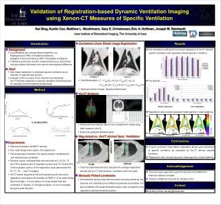
This study evaluates the effectiveness of registration-based dynamic ventilation imaging using Xenon-CT to assess regional ventilation and lung function. By comparing measurements from both registered specific ventilation and Xenon-CT-derived data, we assess accuracy through linear regression and anatomical landmark analysis. Results indicate that inverse consistent linear elastic registration provides a robust estimation of specific ventilation, revealing expansion heterogeneity during tidal breathing. This method offers insights into localized lung function beyond traditional pulmonary measurement techniques, enhancing understanding of diseases like COPD and IPF.

Validation of Registration-based Dynamic Ventilation Imaging
E N D
Presentation Transcript
Validation of Registration-based Dynamic Ventilation Imaging using Xenon-CT Measures of Specific Ventilation Kai Ding, Kunlin Cao, Matthew L. Moehlmann, Gary E. Christensen, Eric A.Hoffman, Joseph M. Reinhardt Iowa Institute of Biomedical Imaging, The University of Iowa Introduction • Consistent Linear Elastic Image Registration • Cost Minimization: • Regional volume change: Jacobian determinant Results • The correlation coefficients from linear regression of Xe-CT derived specific ventilation and registration-based specific ventilation • The landmark error • Registration also reveals expansion heterogeneity in tidal breathing. • Background • Lung diseases can change tissue properties, e.g., Emphysema (COPD): Increased compliance, Idiopathic Pulmonary Fibrosis (IPF): Decreased compliance • Traditional pulmonary function measurements (e.g. spirometry) only give global information and cannot show regional difference. • Goal • Use image registration to estimate regional ventilation as an indicator of regional lung function. • Evaluate of the accuracy of the result by two standards: Xe-CT derived measures of regional ventilation (functional) and 20 manually picked landmarks (anatomical). g h Template, T Target, S Method • Xe-CT Analysis • Observed time-intensity data is fit to compartment model using least squares curve fit. • Pulmonary Analysis Software Suite • Reg.-based vs. Xe-CT derived Spec. Ventilation • Track the deformed slab and calculate the average registration derived and Xe-CT derived Spec. Ventilation within the slab. • Manually Picked Landmarks • 20 landmarks (airway branchpoints) were manually picked by one observer and matched across different pressures and phases. For each landmark, the actual landmark position was compared to the registration-derived landmark position. • Experiments • Siemens Sensation 64 MDCT scanner. • Four adult sheep were used in this experiment. • The sheep were oriented in the supine position, anesthetized and mechanically ventilated. • Dynamic scans: retrospectively reconstructed at 0, 25, 50, 75 and 100% phase points of inspiration portion and 75, 50 and 25% of the expiration portion of the respiratory cycle (denoted as the T0, T1, T2 … and T7 images). • Xe-CT scans: acquired at the end expiratory point during the respiratory cycle (about 45 breaths) at PEEP of 10 for each sheep. Slice thickness = 2.4 mm (about 3.2 times thicker than the volumetric CT slices). 12 contiguous slices = 3 cm of coverage along the axial direction. Before registration After registration 1.13 Apex Expansion Contraction Base 0.88 0%IN-25%IN 25%IN-50%IN 50%IN-75%IN 75%IN-100%IN y Conclusions • Inverse consistent linear elastic registration shows good estimation of specific ventilation as evaluated by Xe-CT derived specific ventilation. • Registration also reveals expansion heterogeneity in tidal breathing. Lung Height x 100%IN 0%IN Acknowledgement • This work was supported in part by the grant HL079406 from National Institute of Health. • Thanks to Matthew Fuld and Dr. Deokiee Chon Contact • Kai Ding, Email: kai-ding@uiowa.edu