Direct Ophthalmoscopy: Examining the Retina and Its Structures
160 likes | 353 Vues
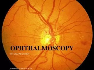
Direct ophthalmoscopy, also known as funduscopy, allows for a detailed examination of the retina and its structures using a specialized instrument. This technique is essential for diagnosing various eye conditions such as glaucoma, retinoblastoma, papilledema, and more. By following specific steps and guidelines, healthcare professionals can assess the optic disc, macula, blood vessels, and other important features of the retina. Understanding the findings can help in early detection and management of eye diseases.

Direct Ophthalmoscopy: Examining the Retina and Its Structures
E N D
Presentation Transcript
Direct Ophthalmoscopy By Thomas Anders Brevik
What is it used for? • Examine the retina and its structures • Also known as funduscopy (examination of the fundus)
Turning the dial to positive (or green) numbers increases the refractive index – short focal length lenses – for examining cornea, iris, or opacities in vitreous or lens. e.g. start at +20 and use the slit light • Turning the dial to negative (or red) numbers decreases – infinite focal length lens that fits your refractive power (individual) – for examining retina, start at +10 as you move in and dim the scope light about halfway • Rule of thumb: You will focus on the retina with same number as your refractive error, then correct for your patients refractive error
Have patient sit in a comfortable position • Tell them to look at something straight ahead and level over your shoulder • Dim light in the room, so patients pupils dilate a little. You can also use mydriatic eyedrops to dilate the pupil • Hold ophthalmoscope in same hand as eye you are looking at, and looking through (e.g. left hand for examining patients left eye, using your left eye) • Hold head steady with thumb above eyebrow, or hold shoulder
At about 30cm distance with light on eye, locate red reflex (seen as an orange glow in the pupil) • Follow red reflex into the eye as 15 degrees lateral to the patients line of vision, this will get you directly into the optic disc • If you cannot find the disc, trace any blood vessels back to it • Examine vessels in all 4 quadrants of eye (upper and lower nasal and temporal quadrants) • Identify macula – slightly darker pigmented area, 2 optic disc widths lateral away from the optic disc • You can tell the patient to look at the light – this will put the macula in your focus, however don’t look at it too long as it can be irritating
Structures of the retina temporal nasal
1 The size, shape and borders of the optic disc • 2 The disc to cup ratio • 3 The relative size of the arteries and veins • 4 The texture of the retina • 5 The color of the retina • 6 Trace the vascular structure to the equator of the retina. • 7 Find the macula and note its color and size
Glaucoma • Identify disc-to-cup ratio • The pink rim of disc contains nerve fibers. The white cup is a pit with no nerve fibers. As glaucoma advances, the cup enlarges until it occupies most of the disc area.
Retinoblastoma • There is a white reflex, rather than red reflex when illuminated • Red reflex is also reduced in cataract
Papilledema • Indicates increased intracranial pressure, e.g. due to hydrocephalus, brain tumor, idiopathic intracranial hypertension or acute intracranial hemorrhage
Proliferative retinopathy and cotton-wool spots Cotton-wool spots are caused by ischemic damage to nerve fibers Compensatory proliferation of vessels Diabetes and hypertension are the main causes
Hypertensive retinopathy • Arteriosclerosis with moderate vascular wall changes (“copper wiring”) to more severe vascular wall hyperplasia and thickening (“silver wiring”) • Arteriovenous crossing abnormalities (arteriovenous nicking) • These vessel changes are better appreciated using the green light (makes the red retina appear in grey tones)
Age-related Macular Degenetation • Wet form: abnormal blood vessel growth w/ hemorrhage and protein leakage • Dry form: Drusen (cellular debris) build-up
http://www.youtube.com/watch?v=AutUi09JIXY&feature=related • http://www.jaapa.com/beyond-the-red-reflex-examining-the-eye-with-an-ophthalmoscope/article/151311/