Download
1 / 38
380 likes | 404 Vues
Explore the types of kinetic motifs in signaling pathways applied to the cell cycle and circadian clocks. Learn about linear response, phosphorylation/dephosphorylation, Michaelis-Menten kinetics, feedback mechanisms, and substrate-depletion oscillations. Understand how toggle switches and negative feedback regulate cell division and control systems. Dive into the dynamic behaviors of molecular signaling cascades in cellular processes.

E N D
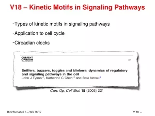
V18 – Kinetic Motifs in Signaling Pathways • Types of kinetic motifs in signaling pathways • Application to cell cycle • Circadian clocks Curr. Op. Cell Biol.15 (2003) 221
Linear Response E.g., protein synthesis and degradation (see lecture V8) S = signal (e.g., concentration of mRNA) R = response (e.g., concentration of a protein) At steady state (which implies S = const): RSS => S RSS linearly dependent on S k0 = 1, k1 = k2 = 2
phosphorylation/dephosphorylation „forward“: R is converted to phosphorylated form RP „backward“: RP can be dephosphorylated again to R S + R => RP with Rtot = R + RP RP => R + T phosphorylated form Find steady state for RP: linear until saturation RPSS Output T proportional to RP level: S Rtot = 1, S0 = 1
E S T kon koff ES Enzyme: Michaelis-Menten-kinetics Reaction rate: Steady state: Total amount of enzyme is constant: => turnover:
The MM-equation Effective turnover according to MM: Pro: • analytical formula for turnover • curve can be easily interpreted: Vmax, KM • enzyme concentration can be ignored Cons: less kinetic information kon, koff, ET => Vmax, KM
Sigmoidal Characteristics with MM kinetics Same topology as before with Michaelis-Menten kinetics for phosphorylation and dephosphorylation. this means that S = Rt - RP KM = R0 Quadratic equation for RP RPSS => sigmoidal characteristics (threshold behavior) often found in signalling cascades S Rt = 10, R0 = RP0 = 1, k1 = k2 = 1
RPSS RPSS RSS S S S Graded Response Linear, hyperbolic, and sigmoidal characteristic give the same steady state response independent of the previous history => no hysteresis BUT: In fast time-dependent scenarios, delay may lead to a modified response
RP(t) t Time-dependent Sigmoidal Response Direct implementation: Parameters: k1 = 1 (mol s)–1, k2 = 1 s–1, R0 = RP0 = 1 mol Initial conditions: R = 10 mol, RP = 0 Time courses for S = 1, 1.5, and 2, RP(0) = 0: equilibrium is reached faster for stronger signal
Adaption - „sniffer“ Linear response modulated by a second species X Steady state: Rss independent of S R changes transiently when S changes, then goes back to its basal level. S X found in smell, vision, chemotaxis, … R Note: response strength ΔR depends on rate of change of S. S => non-monotonous relation for R(S) k1 = 30, k2 = 40, k3 = k4 = 5
Positive Feedback Feedback via R and EP => high levels of R will stay "one-way switch" via bifurcation Found in processes that are "final": frog oocyte maturation, apoptosis, …
Mutual Inhibition - Toggle Switch Sigmoidal "threshold" in E <=> EP leads to bistable response (hysteresis): toggle switch (dt. Kippschalter) Converts continuous external stimulus into two well defined stable states: • lac operon in bacteria • activation of M-phase promoting factor in frog eggs
Negative Feedback S controls the "demand" for R => homeostasis found in biochemical pathways, no transient changes in R for steps in S (cf. "sniffer")
Negative Feedback with Delay Cyclic activation X => YP => RP => X => Oscillations (in a range of S) Proposed mechanism for circadian clocks
Substrate-Depletion Oscillations R is produced in an autocatalytic reaction from X, finally depleting X… Similar to Lotka-Volterra system (autocatalysis for X, too):
M-phase Cell division (cytokinesis) DNA separation (mitosis) cell growth G1-phase DNA replication G2-phase S-phase The Cell Cycle When to take the nextstep???
Simplified Version of Cell Cycle Control System cdc = "cell division cycle“ Cdk1: cyclin dependent kinase 1 Simplification: assume only one type of cyclins (CycB); in reality there are different ones Tyson et al, Curr. Op. Cell Biol.15 (2003) 221
G1 => S — Toggle Switch Mutual inhibition between Cdk1-CycB and CKI (cyclin kinase inhibitor) Degradation of CKI-P Tyson et al, Curr. Op. Cell Biol.15 (2003) 221
Mutual Inhibition ??? Assume: CycB:Cdk1:CKI is stable <=> dissociation is very slow => same topology <=> same bistable behavior (?)
Stoichiometric matrix "(C)" = catalyst R2 A X R1 R3 R4 Rate Equations: Toggle Switch
R1 R2 R3 R5 R4 Rate Equations: G1/S Module R6
R2 R1 R2 A X R1 R3 R3 R5 R4 R4 Comparison: Matrices R6 Difference: catalysts vs. substrates
R1 R2 R2 A X R1 R3 R3 R5 R4 R4 Comparison: Equations R6 Rename species => same rate equations => same behavior
Predicted Behavior: G1 => S Signal: cell growth = concentration of CycB, Cdk1 Response: activity (concentration) of CycB:Cdk1 Toggle switch: => above critical cell size, CycB:Cdk1 activity will switch on Tyson et al, Curr. Op. Cell Biol.15 (2003) 221
G2 => M Dual toggle switch: • mutual activation between CycB:Cdk1 and Cdc25 (phosphatase that activates the dimer) • mutual inhibition between CycB:Cdk1 and Wee1 (kinase that inactivates the dimer) => when the cell grows further during the second gap phase G2, the activity of CycB:Cdk1 will increase by a further step Tyson et al, Curr. Op. Cell Biol.15 (2003) 221
M => G1 Negative feedback loop oscillator i) CycB:Cdk1 activates anaphase promoting complex (APC) ii) APC-P activates Cdc20 iii) Cdc20:APC-P degrades CycB Behavior: at a critical cell size CycB:Cdk1 activity increases and decreases again => at low CycB:Cdk1 level, the G1/S toggle switches off again, => cell cycle completed Tyson et al, Curr. Op. Cell Biol.15 (2003) 221
Overall Behavior Cell divides at size 1.46 => daughters start growing from size 0.73 => switches to replication at size 1.25 G1/S toggle => bistability M/G1 oscillator G2/M toggle => bistability Tyson et al, Curr. Op. Cell Biol.15 (2003) 221
Circadian clocks in mammals and plants Most organisms (animals, plants, fungi and cyanobacteria) enhance their fitness by coordinating their development with daily environmental changes through molecular timekeepers (circadian clocks) Mammals display circadian rhythms in behavioural and physiological processes, such as - sleep - feeding - blood pressure and - metabolism Roles in plants e.g.: - opening of flowers in the morning and their closure at night Circadian rhythms are guided by external light–dark signals that are integrated through intrinsic central and peripheral molecular clocks McClung Plant Cell 18, 792 (2006) 27
Circadian rhythms (1) Circadian rhythms are the subset of biological rhythms with period of 24 h. The term circadian combines the Latin words ‘‘circa’’ (about) and ‘‘dies’’ (day). (2) Circadian rhythms are endogenously generated and self-sustaining. They persist under constant environmental conditions, typically constant light (or dark) and constant temperature. Under these controlled conditions, the free-running period of 24 h is observed. (3) For all circadian rhythms the period remains relatively constant over a range of ambient temperatures. This is thought to be one property of a general mechanism that buffers the clock against changes in cellular metabolism. McClung Plant Cell 18, 792 (2006)
Basic molecular elements of mammalian clocks (a) 2 TFs CLOCK and BMAL1 heterodimerize. (b) BMA1:CLOCK binds to the E-boxes in the promoters of -the PER and CRY genes, - and of clock-controlled genes, and activate their transcription. (c) The translated PER and CRY proteins dimerize in the cytosol, enter the nucleus and inhibit CLOCK-BMAL1–activated transcription. This is the minimal scheme for the mammalian clock. It requires several interconnecting transcriptional, translational and post-translational loops to achieve gene expression with circadian periodicity Sancar, Nat. Struct. Mol. Biol. 15, 23 (2008) 29
Circuit of circadian rhythms in mammals PER: period CRY: cryptochrome Rev-erb, ROR: retinoic acid- related orphan nuclear receptors Ccg: clock-controlled genes CK1: casein kinase; phosphorylates PER and CRY; necessary for their dimerization This step serves to slow down the feed-back cycle. Ko & Takahashi Hum Mol Genet 15, R271 (2006)
Are circadian rhythms relevant for bioinformatics? - RNA-seq and DNA arrays to quantify transcriptomes of 12 mouse organs at 2 hour/6 hour intervals - Circadian genes: defined as genes that oscillate with 24 hour-period (project on sine/cosine functions) Liver contained most circadian genes (-> metabolism), Brain tissue the fewest („the brain never sleeps“)
Globally oscillating genes in mouse tissue Only 10 genes oscillated in all organs: Arntl, Dbp, Nr1d1, Nr1d2, Per1, Per2, and Per3 (core clock factors – as expected), and Usp2, Tsc22d3, and Tspan4. Usp2 - Ubiquitin carboxyl-terminal hydrolase 2 Tsc22d3 - TSC22 domain family protein 3 Tspan4 - The protein encoded by this gene is a member of the transmembrane 4 superfamily, also known as the tetraspanin family.
Overlap of genes/organs (B), how many expected (C)? Extrapolation shows that 55% of all genes are expected to show circadian expression in some organ. Also non-coding RNAs show circadian expression (at lower frequencies). No individual ncRNA oscillated in more than five organs. (ncRNA expression is known to be organ-specific). Conserved ncRNAs means that they are conserved between human and mouse.
(A) Phases + overlap, (B) similarity Time-dependent profiles. Top: all organs Below: individual organs. Most circadian genes show organ-specific expression (small overlap). Peaks often at dawn and dusk. Cluster tissues by similarity of peak phases Tree in panel B shows that developmentally related organs tend to share circadian genes . Venn diagram : organ overlap.
Three Examples (2) Two VEGF-receptors FLT1 and KDR are expressed alternatively. Arrows: times of anti-phasing. (1) Dtx4, a Notch pathway E3 ubiquitin ligase, oscillated in phase with Arntl in all organs (3) IGF1 is most produced in liver -> peaks at the same time throughout body. However PIK3r1 (regulatory subunit for PIK3) peaks at different times in different organs.
Multiple coordinated pathways control PIK3-AKT-MTOR Multiple synchronous (same peak time) receptors feed into PIK3-AKT-MTOR pathway that controls growth and apoptosis. All of them oscillate only in kidney!
Many drug-targets show circadian expression Relevance: drug response will differ significantly depending on day/night time of application Unclear whether these effects are taken into account during clinical studies
Relevance: mouse -> humans, drugs About half of top-100 drugs have half lives < 6 hours!