Download
1 / 25
250 likes | 316 Vues
Explore the intricate molecular pathways involved in muscle breakdown during aging and immobilization in this comprehensive study. Learn about factors influencing skeletal muscle degradation and the activation of various protein degradation systems.

E N D
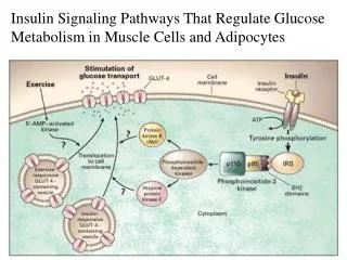
Molecular Mechanisms and Signaling Pathways in Muscle Atrophy in Immobilization and Aging Marina Bar- Shai Abraham Z. Reznick Department of Anatomy and Cell Biology The Bruce Rappaport Faculty of Medicine Technion – Israel Institute of Technology Haifa, Israel
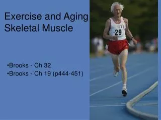
Summary of the main topics • Introduction to aging and muscle protein degradation • In vivo model of immobilization and the stages of skeletal muscle breakdown • In vitro model of the involvement of RNS in activation of NF- κB in muscle cells
The fast phase of muscle breakdown due to immobilization Immobilization (first 24 -48h) Ca+2 influx Increased Ca+2 dependent proteolysis by calpains Initiation of myofibrillar proteins degradation and Z- disk disintegration
The slow phase of muscle breakdown due to immobilization (2-30 days) Infiltration of monocytes and differentiation into macrophages Macrophages activation Synthesis of cytokines IL-1, IL-6, TNF- α by the macrophages Oxidative stress Activation of NF-kB and AP-1 (?) transcription factors Biphasic regulation of the transcription factors by NO: Low levels activate, high levels shut down Upregulation of stress and inflammation genes including iNOS NO, ONOO- RNS Ubiquitin- proteasome- dependent proteolysis Increased muscle wasting Lysosomal proteolysis Ca+2 dependent proteolysis
Mobilization Excessive mobilization (strenuous exercise) Immobilization
In vivo model: Immobilized young and old rats
Experimental design • 6-8 months old female Wistar rats (250-300gr) and 24 months old female Wistar rats (300-350gr) • Immobilization periods : one, two, three and four weeks • Right limbs were immobilized, left limbs served as controls • At the end of each immobilization period the muscles were removed for biochemical and histological studies
Normal vs. immobilized skeletal muscle of an old animal after 4 weeks of immobilization
The activation of various muscle protein degradation systems in immobilized animals
Muscle proteolytic systems Intracellular: • Ca+2– dependent proteases (calpains) • Ubiquitin- proteasome system • Intracellular lysosomal proteases (Cathepsins D, H, L, B., nucleases, lipases, glycosidases, ACP) Extracellular: • Macrophage lysosomal proteases • Matrix Metalloproteases (MMPs): MMP-2, MMP-9
Ubiquitination of muscle proteins following immobilization of young rats Protein staining Immunostaining (anti- Ubiquitin AB.) L-control leg R- immobolized leg
Acid phosphatase activity in normal vs. immobilized (30 days of E.F) muscle of young animals (histochemical staining)
Zymography of gastrocnemius muscles of five young rats after 21 and 30 days of immobilization L-control leg R- immobolized leg
Observations In the slow phase of muscle atrophy due to limb immobilization, the kinetics of activation of the extracellular and the intracellular degradation systems are very similar.
Conclusion There appears to be a link between the activation of the extracellular and intracellular proteolytic systems
The slow phase of muscle breakdown due to immobilization (2-30 days) Infiltration of monocytes and differentiation into macrophages Macrophages activation Synthesis of cytokines IL-1, IL-6, TNF- α by the macrophages Oxidative stress Activation of NF-kB and AP-1 (?) transcription factors Biphasic regulation of the transcription factors by NO: Low levels activate, high levels shut down Upregulation of stress and inflammation genes including iNOS NO, ONOO- RNS Ubiquitin- proteasome- dependent proteolysis Increased muscle wasting Lysosomal proteolysis Ca+2 dependent proteolysis
9th Annual Meeting of The Oxygen Society San Antonio , TX, U.S.A Nov. 20-24, 2002
Acknowledgements • Eli Carmeli, PhD • Raymond Coleman, PhD • Ophir Menashe, MSc • Marina Bar Shai, BSc • Erez Hasnis, BSc • Pessia Shantzer • Bilha Pinkhasi • Shoshan Perek • Yotam Shkedi Thank you for your attention!