Download
1 / 13
140 likes | 317 Vues
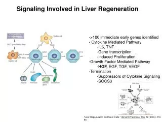
Liver Signaling Pathways. Olga Filippova , Munir Nahri , Akash Patel BMES 471. Methods and Materials. Scaffold Manufacturing. 85:15 and 75:25 PLG microspheres Standard double emulsion process Combined 4:1 with alginate 75:25 PLG encapsulate EGF VEGF and HGF directly into scaffold.

E N D
Liver Signaling Pathways Olga Filippova, MunirNahri, Akash Patel BMES 471
Scaffold Manufacturing • 85:15 and 75:25 PLG microspheres • Standard double emulsion process • Combined 4:1 with alginate • 75:25 PLG encapsulate EGF • VEGF and HGF directly into scaffold 5µm Smith et al, 2004 Lima and Rodrigues, 1999
EGF, HGF Release Kinetics EGF Release from PLG microspheres • 125I labeled EGF • 25±2% retained in micropsheres • 55±3% incorporated into scaffold • Initial 4-day 50% burst • 0.7% per day • Similar HGF analysis Smith et al, 2006
Scaffold Implantation • Three Scaffold Types + Control • Seeded with Lewis rat hepatocytes • 62±5% efficiency • Implanted into SCID mice • 3, 7 or 14 days Experimental Scaffold Setup Control VEGF+EGF VEGF+EGF +HGF VEGF+HGF
Histological Analysis • Vascularization • CD31 antigen • Vascular endothelial cells • Hematocyte Survival • Hematoxylin and eosin • Presence/lack of nucleus • E-800 light microscope
Vascularization Smith et al, 2006 Smith et al, 2004
Hepatocyte Survival Smith et al, 2004 Smith et al, 2006
Hepatocyte Survival • Similar amounts of live cells • More nucleated cells in growth factor scaffold • Less nucleated cells in growth factor scaffold Smith et al, 2006
Hepatocyte Survival Not Improved • Saturation • VEGF - HGF precursor release • HGF - VEGF release • Mesentery vs subcutaneous implantation • Not enough EGF or HGF • Improper release kinetrics Taub, 2004
References • Mohammed, F.F., and Khokha, R. Thinking outside the cell: proteases regulate hepatocyte division. Trends in Cell Biology 15, 555, 2005. • Lima, K.M., and RodriguesJ.M.Jr. Poly-DL-lactide-co-glycolide microspheres as a controlled release antigen delivery system. Braz J Med Biol Res 32, 171, 1999. • Smith, M.K., Peters, M.C., Richardson, T.P., Garbern, J.C., and Mooney, D.J. Locally Enhanced Angiogenesis Promotes Transplanted Cell Survival. Tissue Engineering 10, 63, 2004. • Smith, M.K., Riddle, K.W., and Mooney, D.J. Delivery of Hepatotrophic Factors Fails to Enhance Longer-Term Survival of Subcutaneously Transplanted Hepatocytes. Tissue Engineering 12, 235, 2006. • Taub, R. Liver Regeneration: From Myth to Mechanism. Nature 5, 836, 2004.
Questions? Olga Filippova, MunirNahri, Akash Patel BMES 471