Download
1 / 29
360 likes | 659 Vues
Modeling of Calcium Signaling Pathways. Stefan Schuster and Beate Knoke Dept. of Bioinformatics Friedrich Schiller University Jena Germany. 1. Introduction. Oscillations of intracellular calcium ions are important in signal transduction both in excitable and nonexcitable cells

E N D
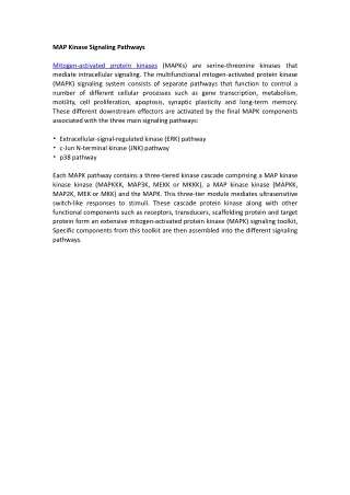
Modeling of Calcium Signaling Pathways Stefan Schuster and Beate Knoke Dept. of Bioinformatics Friedrich Schiller University Jena Germany
1. Introduction • Oscillations of intracellular calcium ions are important in signal transduction both in excitable and nonexcitable cells • A change in agonist (hormone) level can lead to a switch between oscillatory regimes and stationary states digital signal • Moreover, analogue signal encoded in frequency • Amplitude encoding and the importance of the exact time pattern have been discussed; frequency encoding is main paradigm
Ca2+ oscillations in various types of nonexcitable cells Oocytes Hepatocytes Pancreatic acinar cells Astrocytes
Ca2+oscillation Effect 1 Effect 2 Vasopressin Calmodulin Effect 3 Phenylephrine Calpain Caffeine PKC UTP ….. Bow-tie structure of signalling How can one signal transmit several signals?
Scheme of main processes H Caext Y PLC vin R vd vplc PIP2 IP3 vout DAG cytosol + vmo vmi vrel Cam + vserca Cacyt ER mitochondria Caer vb,j proteins Efflux of calcium out of the endoplasmic reticulum is activated by cytosolic calcium = calcium induced calcium release = CICR
Somogyi-Stucki model • Is a minimalist model with only 2 independent variables: Ca2+ in cytosol (S1) and Ca2+ in endoplasmic reticulum (S2) • All rate laws are linear except CICR R. Somogyi and J.W. Stucki, J. Biol. Chem. 266 (1991) 11068
Rate laws of Somogyi-Stucki model H Caext Y PLC v1 R vd vplc PIP2 IP3 v2 DAG cytosol + vmo vmi v5 Influx into the cell: Cacyt=S1 Cam v4 + ER mitochondria Caer=S2 vb,j Efflux out of the cell: v6 proteins Pumping of Ca2+ into ER: Efflux out of ER through channels (CICR): Leak out of the ER:
Temporal behaviour fast movement Relaxation oscillations! slow movement
Many other models… • by A. Goldbeter, G. Dupont, J. Keizer, Y.X. Li, T. Chay etc. • Reviewed, e.g., in Schuster, S., M. Marhl and T. Höfer. Eur. J. Biochem. (2002) 269, 1333-1355 and Falcke, M. Adv. Phys. (2004) 53, 255-440. • Most models are based on calcium-induced calcium release.
2. Bifurcation analysis of two models of calcium oscillations • Biologically relevant bifurcation parameter in Somogyi-Stucki model: rate constant of channel, k5 (CICR), dependent on IP3 • Low k5 : steady state; medium k5: oscillations; high k5: steady state. • Transition points (bifurcations) between these regimes can here be calculated analytically, be equating the trace of the Jacobian matrix with zero.
Usual picture of Hopf bifurcations Supercritical Hopf bifurcation Subcritical Hopf bifurcation unstable limit cycle variable variable stable limit cycle stable limit cycle parameter parameter Hysteresis!
Bifurcation diagram for calcium oscillations From: S. Schuster & M. Marhl, J. Biol. Syst. 9 (2001) 291-314 oscillations Subcritical HB Supercritical HB
Schematic picture of bifurcation diagram variable parameter Bifurcation Very steep increase in amplitude. This is likely to be physiologically advantageous because oscillations start with a distinct amplitude and, thus, misinterpretation of the oscillatory signal is avoided. No hysteresis – signal is unique function of agonist level.
Global bifurcations • Local bifurcations occur when the behaviour near a steady state changes qualitatively • Global bifurcations occur „out of the blue“, by a global change • Prominent example: homoclinic bifurcation
Homoclinic bifurcation Before bifurcation S2 At bifurcation Saddle point Saddle point Unstable focus Homoclinic orbit S1 After bifurcation Necessary condition in 2D systems: at least 2 steady states (in Somogyi-Stucki model, only one steady state) Saddle point Limit cycle
Model including binding of Ca2+ to proteins and effect of ER transmembrane potential H Caext Y PLC vin R vd vplc PIP2 IP3 vout DAG cytosol + vmo vmi vrel Cam + vserca Cacyt ER mitochondria Caer vb,j proteins Marhl, Schuster, Brumen, Heinrich, Biophys. Chem. 63 (1997) 221
System equations 2D model with Nonlinear equation for transmembrane potential DY...
…this gives rise to ahomoclinic bifurcation oscillation Hopf bifn. variable parameter Schuster & Marhl, J. Biol. Syst. 9 (2001) 291 Saddle point As the velocity of the trajectory tends to zero when it approaches the saddle point, the oscillation period becomes arbirtrarily long near the bifurcation.
3. How can one second messenger transmit more than one signal? • One possibility: Bursting oscillations (work with Beate Knoke and Marko Marhl)
Selective activation of protein 1 Prot1 Prot2
Selective activation of protein 2 Prot2 Prot1
Simultaneous up- and downregulation Prot2 Prot1 S. Schuster, B. Knoke, M. Marhl: Differential regulation of proteins by bursting calcium oscillations – A theoretical study. BioSystems 81 (2005) 49-63.
4. Finite calcium oscillations • Of course, in living cells, only a finite number of spikes occur • Question: Is finiteness relevant for protein activation (decoding of calcium oscillations)?
Intermediate velocity of binding is best kon = 500 s-1mM-4 kon = 15 s-1mM-4 kon = 1 s-1mM-4 koff/kon = const. = 0.01 mM4
„Finiteness resonance“ Proteins with different binding properties can be activated selectively. This effect does not occur for infinitely long oscillations. M. Marhl, M. Perc, S. Schuster S. A minimal model for decoding of time-limited Ca(2+) oscillations. Biophys Chem. (2005) Dec 7, Epub ahead of print
5. Discussion • Relatively simple models (e.g. Somogyi-Stucki) can give rise to complex bifurcation behaviour. • Relaxation oscillators allow jump-like increase in amplitude at bifurcations and do not show hysteresis. • At global bifurcations, oscillations start with a finite (often large) amplitude. • Physiologically advantageous because misinterpretation of the oscillatory signal is avoided in the presence of fluctuations.
Discussion (2) • Near homoclinic bifurcations, oscillation period can get arbitrarily high. • This may be relevant for frequency encoding. Frequency can be varied over a wide range. • Bursting oscillations may be relevant for transmitting two signals simultaneously – experimental proof is desirable • Thus, complex oscillations as found in, e.g. hepatocytes, may be of physiological importance • Finite trains of calcium spikes show resonance in protein activation • Thus, selective activation of proteins is enabled
Cooperations • Marko Marhl (University of Maribor, Slovenia) • Thomas Höfer (Humboldt University, Berlin, Germany) • Exchange with Slovenia supported by Research Ministries of both countries.