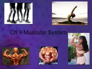
Chapter 9 Muscular System
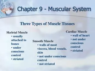
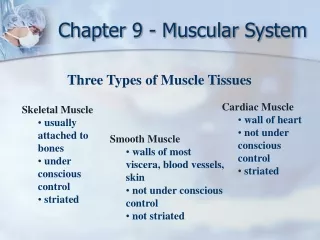
Chapter 9 Muscular System. Three Types of Muscle Tissues. Skeletal Muscle usually attached to bones, skin, deep fascia voluntary striated. Cardiac Muscle wall of heart involuntary striated. Smooth Muscle walls of most viscera, blood vessels, skin involuntary not striated.

Chapter 9 Muscular System
E N D
Presentation Transcript
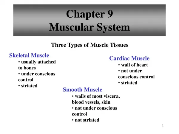
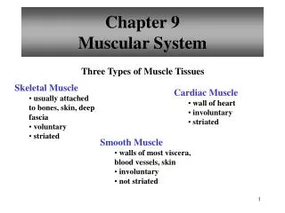
Chapter 9Muscular System Three Types of Muscle Tissues • Skeletal Muscle • usually attached to bones, skin, deep fascia • voluntary • striated • Cardiac Muscle • wall of heart • involuntary • striated • Smooth Muscle • walls of most viscera, blood vessels, skin • involuntary • not striated
Muscle Tissue Characteristics • Excitability • Similar to nervous tissue • Stimulus initiates action potential (impulse) • Contractility • Shorten and thicken • Actively do work when stimulus is received
Muscle Tissue Characteristics • Extensibility • Ability to be stretched • Paired muscle groups • Elasticity • Ability to return to its original shape after contracting or extending
Muscle Tissue Functions • Motion • Maintenance of posture • Heat production • 85% of heat generated from muscles
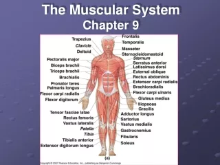
Structure of a Skeletal Muscle • Skeletal Muscle • organ of the muscular system • - skeletal muscle tissue • - nervous tissue • - blood • - connective tissues • fascia • tendons • aponeuroses
Connective Tissue Coverings • Superficial Fascia • Subcutaneous layer • Immediately deep to the skin • Stores fat, insulates, protects, provides pathway for nerves and blood vessels
Connective Tissue Coverings • Deep Fascia • Lines body walls, extremities, and holds muscles together • Splits muscles into functional groups • Pectoralis major/pectoralis minor
Connective Tissue Coverings • Deep Fascia • Functions • Allows free movement of muscles • Fills space • Carries nerve and vascular supply • Sometimes provides origin for muscles
Connective Tissue Coverings • Epimysium • Wraps the entire muscle bundle • Perimysium • Covers muscle fiber bundles (fascicles) • Endomysium • Covers individual fibers within the fascicles • “-mysiums” may extend to become tendon
Epimysium Covers entire muscle Muscles are bundles of fascicles Perimysium Covers individual fascicles Connective Tissue Coverings
Fascicle Many bundles of fibers Fiber Many myofibrils Myofibril Many myofilaments Connective Tissue Coverings
Myofilaments Smallest part of a muscle DO NOT extend entire length of muscle Fit into compartments called sarcomeres Connective Tissue Coverings
Sarcomeres Contraction of muscles occurs here Lie end to end within myofibril Connective Tissue Coverings
Skeletal Muscle Fibers • Sarcolemma • Muscle cell membrane • Sarcoplasm • Muscle cell cytoplasm • Sarcoplasmicreticulum • Muscle cell ER
2 types of myofilaments myosin- THICK contractile protein actin- THIN contractile protein The ability for actin and myosin to change shape allows for the 2 myofilaments to be pulled (slide) over each other Sarcomere Structure
Myofilaments form patterns in the sarcomeres “I” band consist of the disc (stationary) and actin filaments “A” band consist of myosin overlapping with the ends of the actin filaments “H” zone —is within “A” band and contains only myosin Another “I” band Sarcomere Structure
Structures between the disc make up the sarcomere Heads or hooks on the myosin bind with actin to pull the myosin filament over the actin Tropomyosin blocks the binding site on the actin when the muscle is relaxed Sarcomere Structure
Myofilaments • Thin Filaments • composed of actin • associated with troponin and tropomyosin • Thick Filaments • composed of myosin • cross-bridges
Sliding Filament Theory • The myofibril shortens because the myofilaments in each sarcomere slide over each other
Neuromuscular Junction • also known as myoneural junction • site where an axon and muscle fiber meet • motor neuron • motor end plate • synapse • synaptic cleft • synaptic vesicles • neurotransmitters
Motor Unit • single motor neuron • all muscle fibers controlled by motor neuron
Stimulus for Contraction • acetylcholine (ACh) • nerve impulse causes release of ACh from synaptic vesicles • ACh binds to ACh receptors on motor end plate • generates a muscle impulse • muscle impulse eventually reaches sarcoplasmic reticulum
Excitation Contraction Coupling • muscle impulses cause sarcoplasmic reticulum to release calcium ions into cytosol • calcium binds to troponin to change its shape • position of tropomyosin is altered • binding sites on actin are exposed • actin and myosin molecules bind
Sliding Filament Model of Muscle Contraction • When sarcromeres shorten, thick and thin filaments slide past one another • H zones and I bands narrow • Z lines move closer together
Cross-bridge Cycling • myosin cross-bridge attaches to actin binding site • myosin cross-bridge pulls thin filament • ADP and phosphate released from myosin • new ATP binds to myosin • linkage between actin and myosin cross-bridge break • ATP splits • myosin cross-bridge goes back to original position
Relaxation • Acetylcholinesterase • rapidly decomposes Ach remaining in the synapse • Muscle impulse stops • Stimulus to sarcolemma and muscle fiber membrane • ceases • Calcium moves back into sarcoplasmic reticulum • Myosin and actin binding prevented • Tropomyosin slides over binding sites • Muscle fiber relaxes • Sarcomeres return to original length
Energy Sources for Contraction 1) Creatine phosphate 2) Cellular respiration • creatine phosphate – stores energy that quickly converts ADP to ATP
Oxygen Supply and Cellular Respiration • Anaerobic Phase • glycolysis • Occurs in cytoplasm • Produces little ATP • Aerobic Phase • Citric acid cycle • Electron transport chain • Occurs in mitochondria • Produces most ATP • Myoglobin • Pigment that stores extra oxygen
Oxygen Debt Oxygen debt – amount of oxygen needed by liver cells to use the accumulated lactic acid to produce glucose • Oxygen not available • Glycolysis continues • Pyruvic acid converted to lactic acid • Liver converts lactic acid to glucose
Muscle Fatigue • Inability to contract • Commonly caused from • decreased blood flow • ion imbalances across the sarcolemma • accumulation of lactic acid • Cramp – sustained, involuntary muscle contraction
Heat Production • By-product of cellular respiration • Muscle cells are major source of body heat • Blood transports heat throughout body
Muscular Responses • Threshold Stimulus • minimal strength required to cause contraction • Recording a Muscle Contraction • twitch • Single muscle fiber response to an impulse • latent period • Delay between impulse and contraction • period of contraction
Muscular Responses • Period of relaxation • Refractory period • Time when neuron will not respond to stimulus • All-or-none response • Each twitch generates the same force
Summation • process by which individual twitches combine • produces sustained contractions • can lead to tetanic contractions • Lacks partial relaxation
Recruitment of Motor Units • Recruitment • Increase in the number of motor units activated • Whole muscle composed of many motor units • More precise movements are produced with fewer • muscle fibers within a motor unit • Eye has fewer than 10 muscle fibers per motor unit • As intensity of stimulation increases, recruitment of motor units continues until all motor units are activated
Sustained Contractions • Smaller motor units (smaller diameter axons) • Recruited first • Larger motor units (larger diameter axons) • Recruited later • Produce smooth movements • Spinal cord stimulates contractions in different sets of motor units at different times • Muscle tone – continuous state of partial contraction • Maintains posture • Completely lost with loss of consciousness
Types of Contractions • Concentric– shortening contraction • Isotonic – muscle contracts and changes length • Isometric – muscle contracts but does not change length • Eccentric – lengthening contraction
Fast and Slow Twitch Muscle Fibers • Slow-twitch fibers (type I) • Always oxidative • Resistant to fatigue • Red fibers • Contain most myoglobin • Good blood supply • Back muscles • Fast-twitch fatigue-resistant fibers (type IIb) • intermediate fibers • oxidative • intermediate amount of myoglobin • pink to red in color • resistant to fatigue • Limb muscles • Fast-twitch glycolytic fibers • (type IIa) • white fibers (less myoglobin) • poorer blood supply • susceptible to fatigue • Hand muscles, eye muscles
Abnormal Contractions • Spasm • Sudden involuntary contraction of a large group of muscles • Tremor • Involuntary contraction of opposing muscle groups • Fasciculation • Involuntary, brief twitch of a muscle visible under the skin • Occurs irregularly and doesn’t move the affected muscle
Abnormal Contractions • Fibrillation • Similar to fasciculation except it is not visible under the skin • Tic • Twitch made involuntarily by muscles under voluntary control • Eyelids or facial muscles are examples • Generally tics are of psychological origin
Smooth Muscle Fibers • Compared to skeletal muscle fibers • shorter • single, centrally located nucleus • elongated with tapering ends • myofilaments randomly organized • lack striations • lack transverse tubules • sarcoplasmic reticula not well developed
Types of Smooth Muscle • Multiunit Smooth Muscle • less organized • function as separate units • fibers function separately • irises of eye • walls of blood vessels • Visceral Smooth Muscle • single-unit smooth muscle • sheets of muscle fibers • fibers held together by gap junctions • exhibit rhythmicity • exhibit peristalsis • walls of most hollow organs
Smooth Muscle Contraction • Resembles skeletal muscle contraction • interaction between actin and myosin • both use calcium and ATP • both are triggered by membrane impulses • Different from skeletal muscle contraction • smooth muscle lacks troponin • smooth muscle uses calmodulin • two neurotransmitters affect smooth muscle • acetlycholine and norepinephrine • hormones affect smooth muscle • stretching can trigger smooth muscle contraction • smooth muscle slower to contract and relax • smooth muscle more resistant to fatigue • smooth muscle can change length without changing • tautness
Cardiac Muscle • Fibers are quadrangular • Single nucleus • More and larger mitochondria • Contain actin and myosin • Fibers branched and interconnected • 2 separate networks (atria and ventricles) intercalated disc separate each fiber in a network • impulse stimulates the entire network, contraction of the entire network
Cardiac Muscle • atria contract—blood to the ventricle • ventricles contract—blood to the arteries and through the body • auto-rhythmicity—nerve impulses only increase or decrease the rhythmic contractions • remains contracted 10-15 times longer • extra refractory period—allows heart to rest and prevents tetanus
Skeletal Muscle Actions • origin – immovable end • insertion – movable end • prime mover (agonist) – primarily responsible for movement • synergists – assist prime mover • antagonist – resist prime mover’s action and cause movement in the opposite direction
Body Movement • Four Basic Components of Lever • rigid bar – bones • fulcrum – point on which bar moves; joint • object - moved against resistance; weight • force – supplies energy for movement; muscles