Download

1 / 38
430 likes | 2.1k Vues
Radiography of the Shoulder. Jennifer Nicol PGY-1 August 6, 2009. Objectives. BRIEF Anatomy Review Standard shoulder views Radiographs of shoulder injuries NOT: Treatment Other imaging modalities Pediatric imaging. Anatomy. Shoulder Views. Over 15 views of shoulder described

E N D
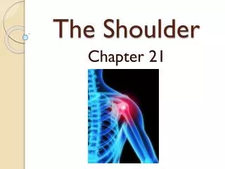
Radiography of the Shoulder Jennifer Nicol PGY-1 August 6, 2009
Objectives • BRIEF Anatomy Review • Standard shoulder views • Radiographs of shoulder injuries • NOT: • Treatment • Other imaging modalities • Pediatric imaging
Shoulder Views • Over 15 views of shoulder described • Trauma series: • 3 views: • AP • Trans-scapular “Y-view” • Axillary • Modified axillary
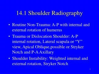
AP view • True AP - 45˚tilt • Glenohumeral joint with no bony overlap • Preferred in trauma • AP int/ext rotation • Highlight tuberosities • Soft tissue injuries • Clavicle and AC joint
Transcapular view • Projects along long axis scapula • Simple, reproducible • Good for visualising anterior, posterior dislocations
Acromion Coracoid Body
Axillary View • Glenohumeral joint in cephalocaudal plane • Lesions of glenoid rim, humeral head, caracoid • Axial view of shoulder
Modified Axillary View • Reverse axillary when pt can’t abduct
Retrospective • 1690 shoulder exams • Mod axillary view used 104 times • Identified additional pathology in 30 cases • No comparison b/t standard and modified axillary
Approach to Shoulder XR • AP: • If ext/int rotation look at tuberosity orientation • Glenohumeral region • Alignment • Distance b/t humeral head and glenoid • Bones • AC region • Other regions (clavicle, ribs, scapular spine,lungs)
Approach to Shoulder XR • Other views: • Humeral head to glenoid • Prox humerus • Glenoid rim • Scapula • Carocoid • Acromion
Type I AC injury • Glenoid • Alignment • Distance • bones • AC • Alignment • Carocoid-clavicle space • Other • Lungs, scapula, ribs, clavicle
Bilateral shoulder dislocation spontaneously reduced with bilateral reverse Hill-Sachs lesions
Posterior Dislocations • Have high suspicion with correct mechanism • Don’t miss – clinical exam important • Look for associated fractures • Types: • Subacromial (98%) • Subglenoid • subspinosus
Bankhart Injury AP Axillary
Anterior Dislocations • 4 Types • Subcoracoid • Subglenoid • Subclavicular • Intrathoracic
Anterior Dislocations • Check Neurovascular exam pre-post reduction • Don’t delay reduction – NV injury increases with time • Recurrence high – 80% <30