Parathyroid Gland Digital Laboratory
280 likes | 662 Vues
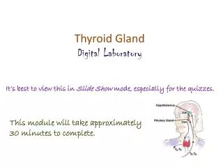
Parathyroid Gland Digital Laboratory. It’s best to view this in Slide Show mode, especially for the quizzes. This module will take approximately 20 minutes to complete. After completing this exercise, you should be able to: identify, at the light microscope level, each of the following:

Parathyroid Gland Digital Laboratory
E N D
Presentation Transcript
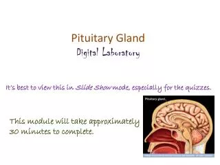
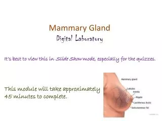
Parathyroid Gland Digital Laboratory It’s best to view this in Slide Show mode, especially for the quizzes. This module will take approximately 20 minutes to complete.
After completing this exercise, you should be able to: • identify, at the light microscope level, each of the following: • Parathyroid gland • Chief cells • Oxyphil cells
PARATHYROID GLAND DEVELOPMENT Usually, there are four parathyroid glands, a superior and inferior on each side of the body and all are histologically similar. Development of the parathyroid glands isn’t that crucial for understanding its histology. Just be aware that these organs develop from the pharyngeal pouches (discussed next year), and migrate into the neck to become buried within the thyroid gland (image C).
PARATHYROID GLAND A low power image of the parathyroid gland reveals two cell types. --Chief (principal) cells are more numerous, smaller, with a slightly eosinophilic cytoplasm. These cells release parathyroid hormone. --Oxyphil cells are less numerous, larger, and have a very eosinophilic cytoplasm due to numerous mitochondria. They are found individually, or clustered in groups. The function of these cells is unknown, yet their presence assists in identifying this organ. The parathyroid gland typically has adipose tissue (AC) interspersed throughout, but this is not too prominent on this image. You probably have come to appreciate that the overall “darkness” of a region on a slide under low power is largely due to nuclear density. This is particularly useful in the parathyroid gland. In the regions where chief (principal) cells are located, the nuclei are closer together, giving an overall “dark” appearance. In regions high in oxyphils, the larger cells mean the nuclei are further apart. This, in combination with the highly eosinophilic cytoplasm, makes those regions of the parathyroid gland “paler”.
PARATHYROID GLAND In this medium powered image of the parathyroid gland, note that the nuclear density is highest in areas of chief cells (CC), and the eosinophilia in the cytoplasm of oxyphil cells (OC). A robust blood supply (BV) is also apparent, as well as adipose cells (A)
PARATHYROID GLAND In this high powered image of the parathyroid gland from our slide set, note that the nuclear density is highest in areas of chief cells (most of the slide), and the eosinophilia in the cytoplasm of oxyphil cells (outlined). There is a binucleateoxyphil cell (common) in the lower outlined region.
PARATHYROID GLAND Video of parathyroid gland – SL126 • Link to SL 126 • Be able to identify: • parathyroid gland • Chief (principal) cells • Oxyphil cells
PARATHYROID GLAND Chief cell Oxyphil cell Oxyphil cells are so full of mitochondria that it’s sometimes difficult to see them individually.
The next set of slides is a quiz for this module. You should review the structures covered in this module, and try to visualize each of these in light and electron micrographs. • Identify, at the light microscope level, each of the following: • Parathyroid gland • Chief cells • Oxyphil cells
Final quiz Self-check: Identify the organ. (advance slide for answers) Adrenal gland
Final quiz Self-check: Identify the organ. (advance slide for answers) Parathyroid gland
Final quiz Self-check: If this image was taken from the pituitary gland, identify cells 8. (advance slide for answers) chromophobes Granules and nuclei in cells at 4 place this image as being from the anterior pituitary.
Final quiz Self-check: Identify the organ. (advance slide for answers) Thyroid gland
Final quiz Self-check: If this image was taken from the adrenal gland, from which part of that gland could this have been obtained. (advance slide for answers) Adrenal medulla
Final quiz Self-check: Identify the outlined region. (advance slide for answers) Zona reticularis
Final quiz Self-check: In this image, #6 are Herring bodies. Identify cells #3. (advance slide for answers) pituicytes Herring bodies are in the posterior pituitary. The majority of nucleated cells here are pituicytesand endothelial cells (2) of blood vessels. Cell bodies of neurons that secrete vasopressin and oxytocin are in the hypothalamus, not the posterior pituitary.
Final quiz Self-check: Identify the organ. Be specific. (advance slide for answers) Posterior pituitary
Final quiz Self-check: Identify the organ. Identify cells 1 (several examples) and 2. Identify 4. (advance slide for answers) Parathyroid gland Cell 1: Chief (principal) cell Cell 2: Oxyphil cell Component 4: Lipid droplet
Final quiz Self-check: Identify the organ. Be specific. (advance slide for answers) Anterior pituitary
Final quiz Self-check: The cell indicated by the arrow could release which hormones? (advance slide for answers) ACTH, FSH, LH, or TSH
Final quiz Self-check: If this image was taken from the adrenal gland, from which part of that gland could this have been obtained. (advance slide for answers) Adrenal cortex
Final quiz Self-check: Identify the outlined region. (advance slide for answers) Adrenal medulla
Final quiz Self-check: Identify the organ. (advance slide for answers) Thyroid gland
Final quiz Self-check: The cell indicated by the arrow could release which hormones. (advance slide for answers) Growth hormone or prolactin
Final quiz Self-check: Identify the cells indicated by the arrows. (advance slide for answers) Oxyphil cells