Download
1 / 18
201 likes | 549 Vues
Congestive Heart Failure Pathophysiology and other relations. Summary of mainly Robbins and Cotran (relevant material from chapter 12). Congestive Heart Failure –CHF.

E N D
Congestive Heart Failure Pathophysiology and other relations Summary of mainly Robbins and Cotran (relevant material from chapter 12)
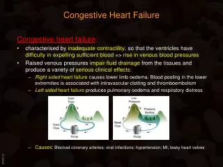
Congestive Heart Failure –CHF Definition: When the heart is unable to pump blood at a rate sufficient to meet the metabolic demands of the tissues or can do so only at an elevated filling pressure. CHF can develop over different time periods: • Chronic work overload (valve disease, HTN or post MI) CHF developing insidiously • Acute hemodynamic stress (fluid overload, acute valvular dysfunction or acute MI) acute CHF
Compensatory Mechanisms during CHF- Cardiac • Frank-Starling mechanism -Normally moderate ventricular dilation during diastole increases the extent of sarcomere shortening and hence inotropy via Frank Starling law… but with further dilation, effective overlap of actin/ myosin filaments is reduced and force of contraction decreases sharply. • Ventricular dilation or hypertrophy - starts off as a compensatory response to increased mechanical work but leads to increased cardiac dysfunction.
Compensatory Mechanisms during CHF- Autonomic Nerves • Increased sympathetic adrenergic activity which increases TPR, HR and inotropy • Reduced vagal activity to heart (athletes have increased vagal tone)
Compensatory Mechanisms during CHF- Hormones • Renin-angiotensin-aldosterone system to increase/ adjust filling volumes and pressures • Natriuretic peptides (ANP/ BNP) to reduce/ adjust filling volumes and pressures
Pathophysiology of CHF – systolic dysfunction Broadly, there are two paths that lead to CHF; the more common one is that of systolic dysfunction (pump failure), where progressive myocardial contractile dysfunction is attributable to: • Ischemic injury, which replaces viable myocytes with fibrous scar tissue. • Pressure - overload (afterload)/ volume – overload (preload) due to valvular (esp. aortic) insufficiency or HTN with resultant hypertrophy. • Dilated cardiomyopathy, with decreased cardiac output related to a weakened and enlarged heart wall. • Drugs causing intracellular damage and oxidative stress. • Defective signal transduction mechanisms eg. Abnormalities in distribution of gap junctions and defects in their proteins may contribute to arrhythmia and heart failure
Pathophysiology of CHF – systolic dysfunction Ventricular end-diastolic pressure and volume increases due to reduced stroke volume transmitted to the atria; • left side congested pulmonary vasculature and oedema. • right side systemic venous hypertension and oedema.
Pathophysiology of CHF – diastolic dysfunction Sometimes however, CHF results from an inability of ventricles to expand when filling with blood during diastole – can’t enrol help of Frank-Starling mechanism. Diastolic dysfunction can occur as a consequence of gross left ventricular hypertrophy, myocardial fibrosis, deposition of amyloid or constrictive pericarditis and normal stiffening with age. The stiffer ventricular wall is unable to allow adequate diastolic filling thus reducing the end diastolic volume which reduces stroke volume and the cardiac output goes down – causing identical symptoms of pulmonary congestion and oedema on the left side and portal hypertension and peripheral oedema on the right; although if systolic function is preserved, diastolic dysfunction may not manifest itself except in physiologic extremes.
Cardiac Hypertrophy and progression to failure The response to increased mechanical work due to pressure or volume overload and other trophic signals cause myocytes to increase in size – producing an increase in the size and weight of the heart. • Increased ventricular systolic pressures (afterload) Concentric hypertrophy (thick wall due to new sarcomeres positioned in parallel with existing ones) • Volume overload (preload) caused by the regurgitant fractions Excentric hypertrophy (dilation due to new sarcomeres positioned in series with existing ones)
Cardiac Hypertrophy and progression to failure Increase in myocyte size and hence metabolic demand is not met with a proportional increase in capillary numbers weak oxygen and nutrient supply causing an hypoxic state, which is especially bad new in systolic dysfunction. This situation means that the hypertrophied heart is vulnerable to decompensation death. So, the compensatory changes in hypertrophied hearts that initially serve to maintain function actually serve to promote heart failure.
Left-sided Heart Failure (LHF) LHF is most often caused by IHD, HTN, aortic and mitral valvular disease and unusual restrictive cardiomyopathies. Morphology and clinical features of LHF are primarily due to pulmonary circulation congestion, stasis of blood on left side of heart, and lack of perfusion to tissues (eg. Kidneys) leading to organ dysfunction.
LHF Morphology • Myocyte hypertrophy and variable interstitial fibrosis microscopically • Myocardial infarction • Left atrial dilation increase risk of AF and hence thrombosis • Perivascular/ interstitial oedema Kerley B lines on x-ray • Widening alveolar septa • Hemosiderin-laden macrophages (HF cells) due to extravasation of RBC’s
LHF Clinical features • Cough • Dyspnea (at rest when condition decompensates) • Orthopnea • CNS hypoxic encephalopathy coma • AF stroke
LHF signs • Tachypea and increased work of breathing • Rales/ Crackles in lung bases • Cyanosis due to severe hypoxia • Laterally placed apical beat • Additional HS (S4 is caused by the atria contracting forcefully in an effort to overcome an abnormally stiff or hypertrophic ventricle causing abnormal turbulent flow) • Murmurs (regurg or stenosis may be cause or result of CHF – soft pan-systolic normally mitral regurgitation during systole – pt should be placed in left lateral position etc)
LHF signs • Decreased CO causes reduction in renal perfusion causing triggering of RAAS – eventually increasing interstitial and intravascular fluid volumes and TPR; this increases organ perfusion and venous return but also exacerbates pulmonary oedema. If renal hypoperfusion worsens azotemia (prerenal). • Left sided Diastolic failure (stiff LV) disables the heart to increase CO in response to increased peripheral demand which exhibits itself most commonly as exertional dyspnea, moreover, increased filling pressures are immediately transmitted back to the pulmonary circulation causing flash pulmonary oedema. Hypertensive females are the most likely to get diastolic LV dysfunction.
Right-sided Heart Failure (RHF) • RHF is most commonly caused by LHF since increase pulmonary pressure is eventually transmitted to the right ventricle. Thus all the causes of LHF are indirect causes of RHF too. • When it does happen though, pure RHF develops from pathology of lungs hence cor pulmonale (heart pathology from lungs). Cor pulmonale usually arises secondary to Interstitial Lung Disease (eg. SLE, RA, TB, sarcoidosis, PCP) but can also arise from pulmonary vasculature disease (PE, pulmonary HTN) or hypoxia-induced pulmonary vasoconstriction.
RHF Morphology and clinical features • Right atrial and ventricular hypertrophy • Portal HTN and associated hepatosplenomegaly and cirrhosis • Pleural, Pericardial and Peritoneal effusions ascites • Peripheral oedema (pitting) and JVPE • More marked azotemia than LHF • CNS hypoxic encephalopathy coma • Parasternal heave due to right sided hypertrophy