Download
1 / 59
600 likes | 921 Vues
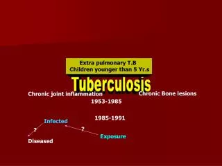
Skeletal Tuberculosis (Part-1) Dr. Sunil Arora Junior Resident Deptt. of Chest &TB Govt. Medical College, Patiala. 1. Skeletal TB accounts for 10 to 35 percent of cases of extrapulmonary tuberculosis.

E N D
Skeletal Tuberculosis (Part-1) Dr. Sunil Arora Junior Resident Deptt. of Chest &TB Govt. Medical College, Patiala. 1
Skeletal TB accounts for 10 to 35 percent of cases of extrapulmonary tuberculosis. • After Lung and Lymph nodes,skeletal TB is the next common type.It constitutes about 1-4% of total TB cases.(More In HIV-infected patients) • Skeletal TB generally occurs due to hematogenous spread from a primary focus. • Coexisting pulmonary TB is seen in appr.50% cases Introduction
Pathogenesis • It produces similar response as in lungs i.e. Chronic granulomatous inflammation.The disease process can start either in bone or in the synovial membrane. • Active focus forms in the metaphysis (in children) or epiphysis (in adults) and the inflammation extends peripherally along the shaft to reach the subperiosteal space. • The inflammatory exudate may extend outward through the soft tissue to form cold abcess and sinuses.The affected bone may undergo fracture.
Metaphyseal infection reaches the joint through subperiosteal space by penetrating the capsular attachment.In adults,the inflammation can spread up to the subchondral area and enter the joint at the periphery where synovium joins the cartilage which leads to the loosening the attachment of articular cartilage and joint displacement. • The epiphyseal plate is not destroyed as the cartilage is resistant to destruction by the TB inflammatory process.
Sometimes the synovium is infected first and the bone is infected secondarily.It is usually in the form of low-grade synovitis with thickening of the synovial membrane and leading to the formation of pannus.Eventually,the articular cartilage is destroyed,joint gets distended with pus,which may burst out to form a cold abcess or discharging sinus.The joint may also get subluxated due to the laxity of the joint capsule and ligaments. • Fibrous ankylosis is a common outcome of healed tuberculosis of the joints except in spine where bony ankylosis follows more often.
Types Two classical forms of disease have been seen;granular and and exudative(caseous) that involve the bone and synovium.Both the patterns have been observed in patients of skeletal TB,one form may predominate. 1.Osseous granular type :- -often follows trauma -insidious onset,constitutional symptoms rare, -soft tissue are slightly warm and tender -healing without residual joint scarring&ankylosis 2. Osseous exudative(caseous) type:- -rapid onset,constitutional symptoms,muscle pain and spasm more marked -soft tissue are warm,swollen and tender -caseous material penetrates into jointdestructive arthritis -healing by joint scarring&ankylosis
Classification • TUBERCULAR SPONDYLITIS- 50% TUBERCULAR VERTEBRAL OSTEOMYELITIS TUBERCULAR DISCITIS • TUBERCULAR ARTHRITIS- 30% HIP JOINT KNEE WRIST JOINT SACROILIAC JOINT ANKLE JOINT AND FOOT SHOULDER JOINT • TUBERCULAR OSTEOMYELITIS- 19% LONG TUBULAR BONES SHORT TUBULAR BONES FLAT BONES- RIBS,STERNUM, SCAPULA, PELVIS • TENOSYNOVITIS/BURSITIS- 1%
Pott’s Spine • Tubercular spondylitis has been documented in ancient mummies from Egypt and Peru and is one of the oldest demonstrated disease.Percival Pott presented the classic description of spinal TB in 1779. • Spinal TB constitutes about 50% of all cases of osteoarticular TB. • MC site: Lower thoracic and lumber region followed by middle thoracic and cervical vertebrae.
Regional Distribution • 1 Cervical 12% • 2 Cervicodorsal 5% • 3 Dorsal 42% • 4 Dorsolumbar 12% • 5 Lumbar 26% • 6 Lumbosacral 3%
Anatomy • Vertebre develops from the sclerotome on either side of notochord • Each pair of sclerotome (common blood supply) form Lower half of one vertebra and upper half of one below it along with intervening disc. • Therefore ,infections via the arteries involve the embryological section.
Patterns of Vertebral Involvement Four patterns : • Paradiscal • Central • Anterior • Appendiceal (Posterior)
Paradiscal • Commonest type • Spread through arterial supply • Bacteria lodge in the contiguous areas of two adjacent vertebrae granulomatous inflammation leading to erosion of vertebral margins loss of nutrition of intervertebral disc Disc degeneration • When the intervertebral discs have been completely destroyed,the adjacent bodies fuse with each other.
Central Lesions • Body of single vertebra is affected. • Starts in the centre of the vertebral body. • Infection at this site probably reaches through Batson’s venous or branches of post.vertebral artry. • Lytic area develops in the centre of vertebral body leading to balooning of vertebral body mimicking tumour • Later stages-concentric collapse resembling Verebra Plana. • Disc space is not/minimally affected
Anterior lesions— Infection starts in the anterior part of vertebral body and spreads under the ant. Longitudinal ligament. • Post/Appendiceal— Pedicle,lamina,spinous process or transverse process of vertebra are affected.
Clinical Features • Constitutional symptoms,such as fever, night sweats,loss of weight and appetite may occur before symptoms related to spine. 1. Pain-can be Localised to the site(MC early symptoms) Radicular worsen with activity and at night(night cries) 2. Stiffness-Protective mechanism of body where paravertebral muscle go into spasm to prevent movement at the affected vertebra.
3. Cold abcess- Patient may present the first time with swelling(cold abcess) or due to its compression effects:- Retropharyngeal abscess --Dysphagia,dyspnea, Hoarseness of voice Mediastinal abscess --Dysphagia Psoas abscess -- Flexion deformity of hip -No usual signs of inflammation like heat ,redness etc. -Follows paths of least resistance along facial planes,blood vassels &nerves.
Presentation of cold abscesses from different regions of spine • Cervical spine- Exudate collects behind prevertebral facia and may protrude as Retropharyngeal abscess, It may track down in mediastinum to enter into trachea,esophagus or pleural cavity.It may spread lateraly into sterno-cleido mastoid and form abscess in neck. • Thoracic spine- It may confined locally and may appear on X-ray as fusiform or bulbous paravertebral abscess .It can compress spinal cord or penetrate the ant.longitudinal ligament to form a mediastinal abscess or pass downward through medial arcuate ligament to form lumber abcess. • Lumber spine- Most commonly enters the psoas sheathPsoas abscess,also abscess in scarpa’s triangle,medial aspect of thigh
4.Fallacious history of trauma- Trauma may draw attention to a pre-existing lesion or may activate a latent tubercular focus 5. Paraplegia-Rarely it is the presenting symptom. 6. Wedging :- Dorsal spine : Line of weight bearing passes ant to vertebrae.Ant wedging occurs.In late stages leading to kyphotic deformity Cervical and lumber spine : Wedging is less due lordotic curvature 7. Gibbus-If patient presents late
Examination • Gait- Patient walks with short steps to avoid jerking the spine.In TB of cervical spine,patient often supports his head with both hands under the chin and twists his whole body in order to look sideways. • Attitude and deformity :- Cervical spine : Stiff,straight neck Thoracic spine : Kyphus or gibbus,walks very carefully Lumber spine : Loss of lumber lordosis
3. Paravertebral swelling- Superficial cold abcess may present as fullness or swelling on the back,along the chest wall, usually fluctuant. It is important to look for cold abcesses in not so obvious locations,depending upon the region of spine involved. 4. Neurological Examination-To determine if there is any neurological compression and to determine level and severity of neurological compression 5. General examination- For any active or healed lesion,for any other systemic illnesses like,diabetes,HT,jaundice etc.
Investigation • Radiological examination :- 1. Xray spine-AP,Lateral 2. CXR-for primary focus 3. Xray abd-KUB,if psoas abcess is suspected or to find out Primary in abd. • The classic roentgen triad in spinal tuberculosis is primary vertebral lesion, disc space narrowing and paravertebral abscess. • On an avg. 2.5 to 3.8 vertebrae are involved
Paradiscal : Reduction in disc space- Initialy there is demineralization with indistinct bony margins-gradually disc space narrowing occurs.The disc space may eventualy disappear leading to wedging. • Lateral X-ray is better for evaluation of disc space. • Takes 3-5 months for bony destruction to become visible on X-ray • More than 30 % of mineral must be removed from bone for a radiolucent lesion to be visible
Radiological Examination 2. Central : Lytic area in the centre of vertebral body which enlarges and baloons out like tumour.Disc space is preserved. 3. Anterior : Shallow excavation on anterior or lateral surface of vertebral body. 4.TB of posterior elements is usually not detected in early stages in radiographs. Late Stages --Kyphotic deformity,lateral shift and scoliosis,if one side of vertebrae is completely destroyed Hemivertebrae Signs of healing—bone density improves,sclerosis, fusion of contiguous vertebrae. Skip lesions as involvement of non contiguous vertebrae (7 – 10 % cases).
Tuberculous spondylitis. Lateral radiograph demonstrates obliteration of the disk space (straight arrow) with destruction of the adjacent end plates (curved arrow) and anterior wedging.
There is narrowing of the disk space at L4-5, with end plates indistinctly outlined. CT section through the disk space clearly shows destructive changes of the disk and vertebral end plate characteristic of infection
X-ray dorsolumbar spine showing vertebra plana of T10 vertebra. Disc space is well maintained.
Subligamentous spread of spinal tuberculosis. Lateral radiograph demonstrates erosion of the anterior margin of the vertebral body (arrow) caused by an adjacent soft-tissue abscess.
Destruction of the right side of the vertebral body and the neural arch, with the remainder of the body maintaining its shape. The lower disc space is narrowed on the right side; the upper space is almost normal and there is a small paravertebral abscess.
Evidence of cold abcess on X-rays Paravertebralabcess : Paravertebral soft tissue shadow corresponding to the site of affected vertebrae in AP view can • Fusiform [bird nest abcess] : L>W,seen in dorsal spine area. • Globular or tense : W>L,pus under pressure a/w paraplegia • Widened mediastinum : Abscess from dorsal spine may present as widened mediastinum
Aneurysmal phenomena : Concave erosions along the margins of vertebral bodies produced by long standing tense paravertebral abcess,usually in dorsal spine • Retropharyngeal abcess : In cervical spine TB,seen on lateral view : increase in soft tissue thickness (>4mm) in front of C3 vertebral body. • Psoas Abcess : In dorso-lumber and lumber TB,psoas shadow on X-ray of abd may show a bulge.
CT Scan • CT demonstrates abnormalities earlier than plain radiography. It is of great value in the demonstration small paravertebral abscess,not otherwise seen on plain X-ray or any calcification within the cold abscess or visualizing epidural lesions containing bone fragments.
A CT scan showing destruction of the neural arch on both sides, as well as of the vertebral body. Arrows, anterior spinal abscess
Tuberculous spondylitis. Axial CT scan demonstrates lytic destruction of the vertebral body (black arrow) with an adjoining soft-tissue abscess (white arrow). Calcified psoas abscess. Axial CT scan demonstrates bilateral tuberculous psoas abscesses with peripheral calcification (arrows).
MRI • Investigation of choice to evaluate the type and extent of compression of cord,to know the spread of disease under the anterior or post.ligament, most effective to demonstrate neural compression,helps to differentiate between TB and pyogenic infection :- TB – Thin and smooth enhancement of the abcess wall Pyogenic – Thick and irregular • MRI is more sensitive than x-ray and more specific than CT in the diagnosis of spinal tuberculosis.
Cord changes • Conventional radiograph-no information • CT –inadequate assesment • MRI -gives invaluable information Cord oedema or focal myelomalacia is seen as hyperintense signal and It can also diagnose extraosseous extradural granuloma.
‘Gibbus formation’ in the thoraco-lumbar region of a patient with spinal tuberculosis (left). The magnetic resonance shows spinal tuberculosis at T10–T12. Spinal tuberculosis causes the destruction, collapse of vertebrae and angulation of vertebral column
X-ray of cervical region which shows spinal tuberculosis of cervical six to seven vertebrae and a retropharyngeal abscess (left). T1-weighted image of an MRI of same patient, which shows destruction of C6–C7 vertebrae
Myelography • To determine the level of obstruction • May be indicated in cases with ‘spinal tumour syndrom’ • In cases of multiple vertebral lesion • When pt has not recovered after decompression
FNAC : Especially of cold abcess,ZN Stain,C/S • Biopsy : May be required in cases of doubtful diagnosis • Other Investigation : To support the diag:- Increased ESR,Decreased Hb,relative lymphocytosis,Mantoux
Differential Diagnosis • Congenital defects like Schmorl’s disease, Scheurermann’s disease. • Infetious conditions like Acute pyogenic,Typhoid spine,Brucella spondylitis,Mycotic Spondylitis,Syphillis • Tumours Conditions :- Benign : Hemangioma,Giant cell tumour,Aneurysmal bone cyst. Malignant : Ewing’s sarcoma,Osteogenic sarcoma,Multiple myeloma,secondaries • Traumatic conditions
Treatment • Before availability of ATT,mortality rate was 30 % or severe crippling deformities • Aim of treatment is to achieve healing of disease & to prevent,detect early and promptly any complication like paraplegia • Rest: Bed rest for pain relief and to prevent further collapse and dislocation of diseased vertebrae.in children body cast is used.For cervical spineMinerva jacket&coller
Building up of patient’s resistance : High protein diet. • ATT : This remains the cornstone of management, completed by rest,nutritional support and splinting, as necessary.However, there is difference of opinion reg.the duration of drug therapy.Short course chemotherapy for nine months has shown good results in patients with disease coused by succeptible microorganisms. • Antibiotics : For persistently draining sinuses which get secondary infection. • Bed soar care and to treat other comorbid conditions.
Mobilisation : Gradual as improvement begins sit & walk,the spine is supported with coller(cervical),brace (dorso-lumber spine) • Cold abcesses may subside with ATT,if present superficially may need aspration(antigravity insertion of needle through a zig-zag tract) or evacuation(wound closed without a drain) • Sinuses: Mostly heal within 6-12 weeks.If no improvement Excision of tract
Indication for surgery 1. Doubtful diagnosis where open biopsy is necessary 2. Failure to respond to ATT 3. Radiological evidence of progression of bony lesion or paraspinal abcess shadow. 4. Imminent vertebral collapse. 5. Instability of spine and subluxation or dislocation of vertebral body.
Procedures • Anteriolateral decompression with interbody bone grafting.Grafts placed anteriorly. • Costo transversectomy with dempression • Metallic implants& titanium cage filled with cancellous bone when whole body is destroyed. • Kyphotic deformity is prevented by ant debridement,ant inerbody fusion&post fusion.
Pott’s Paraplegia • It is a most serious complication of spinal TB,incidence is appr 20%. • MC in dorsal spine because it is the narrowest region,abcess remains confined under tension and even a small compromise can lead to neurological deficit,infection is common in this area and spinal cord terminates below L1