Advancements in High-Resolution PET for Prostate Imaging: New Probes and Performance Insights
This research presents innovative developments in high-resolution positron emission tomography (PET) specifically designed for prostate cancer imaging. We address the challenges of imaging small prostatic lesions by utilizing advanced detector technologies and mechanical designs aimed at enhancing probe sensitivity and performance. Our findings highlight significant improvements in resolution and signal detection, with Monte Carlo simulations indicating enhanced coincidence event detection. This work integrates insights from multiple institutions and aims to optimize PET instrumentation for better clinical outcomes in prostate cancer diagnostics.

Advancements in High-Resolution PET for Prostate Imaging: New Probes and Performance Insights
E N D
Presentation Transcript
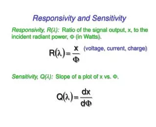
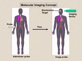
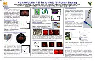
P21-20 30mm 20mm 45mm 40mm 85mm Conventional PET ring Excellent resolution close to probe Modest resolution PET ring High resolution endorectal PET detector (“probe”) Object position External probe insert High Resolution PET Instruments for Prostate ImagingNeal Clinthorne1, Stan Majewski2, Karol Brzezinski3, Sam S. Huh4, Jessie Carr4, Zhewei Chen4, Emma Salomonsson4, Aashay Yande41Dept. Radiology University of Michigan, Ann Arbor, MI 2Center for Advanced Imaging, Dept. Radiology, West Virginia University, Morgantown, WV 3IFIC/CSIC University of Valencia, Spain, 4Dept. Biomedical Engineering, University of Michigan, Ann Arbor, MI Background Prototype Development Probe Sensitivity and Anticipated Performance As an imaging modality in prostate cancer, positron emission tomography (PET) allows the possibility of “engineering” radiotracers to follow specific metabolic pathways or indicate the presence of biomarkers associated with disease. However, our considerable investment in tracer development cannot be used to full advantage if PET instrumentation is not capable of imaging small prostatic lesions well. Presently, PET can detect prostatic lesions of ~8mm diameter. This due to strong attenuation of the annihilation radiation from the prostate, intrinsic resolution limits of the scanner, and patient motion during scanning. Although the technologies associated with the basic concept and detector are absolutely essential, there are many additional ingredients necessary for testing with human subjects. These include the mechanical design of the probe housing, considerations for temperature control, mounting of electronics, and tracking the position and orientation of the probe, which may move during the PET acquisition. To address these issues, we enlisted a team from a two-semester design course in the Department of Biomedical Engineering at Michigan. Shown immediately to the right is the current design, which houses the detector, motion sensors, and EM shielding (see author for 3D model). Monte Carlo simulations using an anthropormorphic phantom, probe, and 80cm dia. PET ring showed that ~12–60% more coincidence events from the prostate were detected depending on detector size (upper right). Plot below right shows effect on performance over PET ring alone in terms of noise advantage vs. resolution for only 12% additional efficiency and for probes having 1mm, 1.5mm, and 2mm resolution. A noise advantage of 10 is equivalent to 102 or 100x the number of events. C-11-choline PET image of prostate cancer. Resolution v. Noise Tradeoff One way to improve resolution is to use an image reconstruction method that models—and then undoes—blurring inherent in the measurements. While this can work to some extent, it inevitably increases the level of noise in reconstructed images. There is an intrinsic “resolution-noise” tradeoff associated with each imaging system. Shown at right are resolution-noise tradeoff curves for three PET scanners having different intrinsic resolution (4mm, 6mm, and 8mm FWHM). Notice how quickly the noise level rises as one attempts to work at resolution better than supported by the intrinsic resolution. Mechanical Testing Detector Technology Originally, aluminum was the material of choice for constructing prototypes; however, it was determined that ABS plastic was considerably less expensive and had the required strength. (Yield strength 42 MPa, highest stress is ~22 MPa.) Seams will be sealed with parylene, which is biocompatible and a good electrical and moisture barrier. New silicon photomultipliers and arrays of LYSO scintillators are enabling technologies for constructing miniature, high-performance PET detectors. Shown at right is a diagram of a probe detector having depth-of-interaction resolution developed by our collaborators at West Virginia University. For details on the detectors, please visit poster P21-17 Demonstration of High Resolution PET Concept Motion Tracking Tests Better Resolution Through Better Measurements The basic high resolution PET concept has been demonstrated with a partial BGO PET ring (100cm dia.) and two high resolution silicon detectors (1.4mm res.) located close to a small (4cm) field-of-view. Reconstructed resolution phantom images shown are from BGO-BGO, BGO-Si, and Si-Si events (left-to-right). Spot diameters are 4.8, 4.0, 3.2, 2.4, 1.6, & 1.2mm. Note the “lever-arm” resolution improvement over BGO-BGO reconstruction with BGO-Si data (as for the prostate probe). Demonstration of the “Limited-Angle” Imaging Probe Low-noise and accurate position-tracking is essential for maintaining high spatial resolution. Shown above right is a setup used for measuring linearity and resolution of the Ascension 3D Trakstar pulsed magnetic system as well as position, linearity, and residual plots. Range measured is considerably larger than will be needed in practice. Resolution (rms) is ~0.3mm, which will not significantly degrade performance of probe. Another method for improving spatial resolution is to use a high-resolution detector probe as an add-on to a standard PET scanner. While “ring-ring” coincidences will still support modest resolution, “probe-ring” coincidences will have excellent resolution near the high resolution probe detector. Moreover, effects induced by positron acolinearity, photon depth-of-interaction, and patient motion are significantly reduced for activity within the prostate. Photon attenuation is also reduced. Shown at right is a plot of the intrinsic spatial resolution as a function of distance from the probe for a conventional PET ring having 6mm intrinsic resolution and probes having 1mm, 2mm, and 3mm intrinsic resolution. Note that the improvement can be significant even at >5cm from the probe face. Conclusions The panel above showed use of full-angle tomographic data while the prostate imaging probe will only acquire limited-angle high-resolution data. To evaluate effects, an external probe acquiring limited-angle data was simulated and results are shown above. External ring had 4mm and probe had 1mm resolution. Images left-to-right are preliminary reconstructions from ring-ring, probe-ring, and probe-ring and ring-ring coincidences combined. Combined performance should exceed that of probe-ring alone as work progresses. Images at left show effects of adding limited angle BGO-Si data to poorer resolution BGO-BGO data alone (from above testbed). Although artifacts are present, resolution improvement is noticeable. Work to develop a high resolution prostate imaging add-on is proceeding rapidly in parallel along three fronts: detector design, probe theory and image reconstruction, and housing design for testing with human subjects. Progress toward our goal of better PET imaging of the prostate has been significant with development of several suitable high-resolution detectors, demonstration of the high resolution PET and PET imaging probe concepts, and design of a prototype housing suitable both for initial tests with phantoms and for eventual testing with prostate cancer patients. Work remains on interfacing the probe to a clinical PET/CT instrument; however, PET manufacturers have shown interest in the technology, which should be useful for guiding biopsy (especially when combined wth MRI or ultrasound images). This work was partially supported by the U.S. Army Medical Research and Materiel Command CDMRP Prostate Synergistic Idea grant W81XWH-09-1-0420.