Download
1 / 20
210 likes | 733 Vues
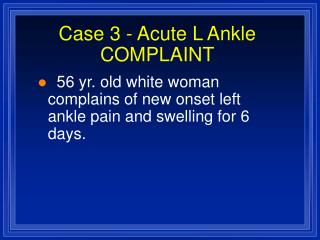
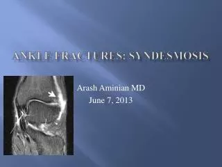
Ankle Fractures: syndesmosis. Arash Aminian MD June 7, 2013. DISCLOSURE. I have no conflicts of interest. Syndesmosis : Why?. 20% of all ankle sprains 10% of all ankle fractures. Questions: evidence vs eminence based. A deltoid tear can be seen with medial malleolar fracture? T

E N D
Ankle Fractures: syndesmosis Arash Aminian MD June 7, 2013
DISCLOSURE • I have no conflicts of interest
Syndesmosis: Why? 20% of all ankle sprains 10% of all ankle fractures
Questions: evidence vs eminence based A deltoid tear can be seen with medial malleolar fracture? T Medial tenderness is predictive of deep deltoid tear? F Intra-operative post reduction stress views should be performed after lateral malleolus fixation? T
Know your enemy Bone quality Pre-reduction films: energy of injury Patient factors: BMI Commonly mal-reduced: IR or anterior translation Not every fracture is the same Don’t be in a hurry
Anatomy: Tib fib syndesmosis AITF PITF Transverse TF IOS
Anatomy: ligamentous stability Deltoid Ligament: Superficial Deep: prevents lateral shift and resists ER
Vascular Anatomy of the tibiofibularsyndesmosis Contribution from peroneal/AT Peroneal artery perforates IO membrane 3 cm proximal to ankle JBJS 2012
Biomechanics Fibula migrates 2.4 mm and ER with DF Deltoid cut 2 mm shift/15 degrees valgus – Harper 1987
LOCATION OF FRACTURE AND SYNDEMOTIC INJURY • Can have a high fibula fx without complete rupture of the IO, deep deltoid • 39% Weber B – Tornetta JOT 2007 Boden JBJS 1989
Syndesmosis fixation? Why, When, How??? Maintain talus in mortise 1 mm shift-42% loss of contact surface RH 1976, 40% Lloyd FAI 2006 2 mm – 49% pressure Zindrick 1985
Clinical evaluation Medial Tenderness Anterior tenderness – nonspecific ER test Squeeze test Check proximal fibula tenderness
Radiographic evaluation AP WB x-rays Mortise: Medial clear space “Shenton’s line” Talocural angle 83 +/- 4 degrees on mortise Tib fib overlap/clear space
Syndesmosis classification Stable vs Unstable Acute vs Chronic Latent vs Frank
Hook test sensitivity/specificity 0.25/0.98 ER test sensitivity/specificity 0.58/0.96
Fixation 3 vs 4 cortices? 3.5 vs 4.5 mm? Biomechanically similar (FAI 2000) Location of the screw? Postero-lateral to anteromedial TSS fixation level above plafond? 2-5 cm
FIXATION • Use a clamp? Prevents motion, shifting of the drill hole • Position of the ankle? DF (JBJS 2001) • HWR? • Fix a high fibula fracture vs only screws? • Fixing the posterior malleolus? Displacement/stability
Post-op rehab 2 wks in splint, then CAM NWB 6 wks WBAT with boot for 6 wks Counsel about hardware breakage