Download
1 / 21
220 likes | 677 Vues
Foot Stress Fractures. Rachel Callis February 2, 2006 Radiology. Anatomy. Divisions Forefoot toes, metatarsals Midfoot 3 cuneiforms, cuboid, navicular Rearfoot talus, calcaneus Ankle tibia, fibula. Anatomy. Stress Fractures. Originally described in 1855

E N D
Foot Stress Fractures Rachel Callis February 2, 2006 Radiology
Anatomy • Divisions • Forefoot • toes, metatarsals • Midfoot • 3 cuneiforms, cuboid, navicular • Rearfoot • talus, calcaneus • Ankle • tibia, fibula
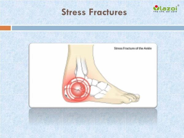
Stress Fractures • Originally described in 1855 • Due to repetitive stress, bone fatigue • Most common in LE • “march fracture,” “en pointe” • 10% of pts in sports medicine practice • 17% bilateral
Stress Fractures • 34% Tibia • 24% Fibula • 18% Metatarsals - 2nd and 3rd metatarsals • Navicular most common of tarsal bones • Calcaneous, the rest
Foot Radiograph ABC’s • A • Alignment • TMTJs = tarsometatarsal joints • B • Bones • Trace the cortex = AP medial 2, MO lateral 3 • Accessory bones • C • Congruity • Forefoot joints on AP and MO
Fracture Classification • Salter-Harris
Stress Fractures • Injuries • Acute < 2 wks • Subacute 2-6 wks • Chronic 6+ wks • Fractures usually linear • Located in central 1/3
Stress Fractures • Diagnosis • Discrete localized swelling and dull pain • Worsens with exercise or weight bearing • Tenderness to palpation at injury site • Continuum • X-rays • may not appear for 2-10 wks • Sensitivity as low as 15% • 50% of repeated x-rays will show fractures
Radiographs Initial 2 wks 1 mo 3 mo
Stress Fractures • Triple phase nuclear bone scans more sensitive • MRI/CT
Figure 5. Athlete with complete navicular stress fracture. A, Radiograph with complete navicular stress fracture (arrow). B, CT scan of same athlete more clearly demonstrating navicular stress fracture (arrow). Navicular Stress Fracture
Treatment • RICE • Rest • Ice (limit to 20 min/hr) • Compression • Elevation • NSAIDs • Short leg casts for metatarsal and navicular fractures
Prevention • Gradual increase in exercise intensity • Stretching • Supportive shoes
Works Cited • Clanton, TO, DA Porter. Primary care of the injured athlete, Part 1: Primary care of foot and ankle injuries in the athlete. Clin Sports Med. 1997. 16(3):435-66. • Damon, JS, AH Newberg. Imaging of stress fractures in the athlete. Radiol Clin N Am. 2002; 40:313-331. • Gellman, R, S Burns. Walking aches and Running Pains. Orthopedics. 1996. 23(2):263-280. • Hatch, RL, S Hacking. Evaluation and management of toe fractures. Am Fam Physician. 2003. 68(12):2413-2418. • Judd, DB, DH Kim. Foot fractures frequently misdiagnosed as ankle sprains. Am Fam Physician. 2002. 66(5):785-794. • Mittlmeier, T, P Haar. Sesamoid and toe fractures. Injury, Int J Care Injured. 2004. 35:S-B87-S-B97. • Pearse, EO, B Klass, SP Bendall. The ‘ABC’ of examining foot radiographs. Ann R Coll Surg Engl. 2005. 87:449-451. • Ribbans, WJ, R Natarajan, S Alavala. Pediatric Foot Fractures. Clin Orthop. 2005. 432:107-115. • Sanderlin, BW, RF Raspa. Common stress fractures. Am Fam Physician. 2003; 68(8):1527-1532.